This decision memorandum does not constitute a national coverage determination (NCD). It states CMS's intent to issue an NCD. Prior to any new or modified policy taking effect, CMS must first issue a manual instruction giving specific directions to our claims-processing contractors. That manual issuance, which includes an effective date, is the NCD. If appropriate, the Agency must also change billing and claims processing systems and issue related instructions to allow for payment. The NCD will be published in the Medicare Coverage Issues Manual. Policy changes become effective as of the date listed in the transmittal that announces the Coverage Issues Manual revision.
To:		File: FDG Positron Emission Tomography (PET) CAG-00065N

From:	Sean Tunis, MD, MSc
		Director, Coverage and Analysis Group

		Mary Stojak
		Health Insurance Specialist, Coverage and Analysis Group

		Samantha Richardson
		Health Insurance Specialist, Coverage and Analysis Group

		Mitchell Burken, MD
		Medical Officer, Coverage and Analysis Group

		Michael Londner, MD, MPH
		Medical Officer, Coverage and Analysis Group

		Madeline Ulrich, MD
		Medical Officer, Coverage and Analysis Group

		Shana Olshan
		Technical Advisor, Coverage and Analysis Group

Subject:		National Medicare Coverage Decision on FDG PET

Date:		December 15, 2000
In this memorandum we: 1) describe FDG PET scans; 2) review the history of Medicare's coverage policy on PET scans and give an explanation of the coverage guidelines; 3) present and analyze the relevant scientific data including the literature submitted by the requestor; and 4) delineate the changed national coverage policy and HCFA’s reason for the coverage decision policy. A summary of the new coverage policy can be found in the last section of this document prior to the appendices.
Description and Background of FDG Positron Emission Tomography (PET)
PET is a noninvasive diagnostic imaging procedure that assesses the level of metabolic activity and perfusion in various organ systems of the human body. Images are obtained from positron-emitting radioactive tracer substances (radiopharmaceuticals) that are usually administered intravenously to the patient
Positron-emitting radioisotopes were first discovered in the 1930’s. FDG PET has been evaluated for several decades in pre-clinical models, and is premised on basic research in biochemisty and biology that have established the basis of glucose metabolism in normal cell function, and it’s alteration in diseases like cancer, ischemic heart disease and some neurological disorders. The first PET scanners were developed in the United States in the 1970’s with the first scan of a human reported in 1978. Through the early 1980's, PET scans were used primarily in research and predominantly focused on the neurosciences because scanners were typically only large enough for head studies. Due largely to the emergence of two major commercial suppliers in the mid-1980’s, PET scanners have become capable of whole body imaging and increased computer processing capability. Improvements in the technology have had a significant impact on the quality of PET’s image reconstruction and display.
PET’s Ability to Identify Pathophysiology
Most of the disease-specific indications addressed in this coverage determination are related to PET use for various types of malignancies. As a group many of these diseases, which frequently are life-threatening, involve uncontrolled reproduction and spread of abnormal malignant cells. In adults, normal cells in most tissues divide only infrequently to replace worn-out or dying cells and to repair injuries. Malignant cells, which are both structurally and functionally abnormal, compete with and destroy normal cells and may spread throughout the body. They may aggregate in solid masses referred to as tumors. The spread of malignancy to a new site is called metastasis.
Classification of cancer by its appearance under a microscope and the part of the body in which it began, is important because different types of cancer vary in growth rates; how they spread through out the body, and in their susceptibility to various anticancer therapies. An accurate diagnosis of where the cancer originated in the body and its type is necessary so that the physician can determine the appropriate clinical management of the patient.
As a molecular diagnostic imaging modality, PET can detect rates of biological activity, as contrasted other imaging modalities such as x-ray films, computed tomography (CT), and magnetic resonance imaging (MRI), which depict the anatomical location of both normal and abnormal structures in the body. Malignancies can cause abnormalities of blood flow or metabolism before anatomic changes are apparent. Thus, disease can be detected by PET when anatomic imaging studies are still normal, and may be informative in differentiating benign from malignant processes. PET evaluation of tissue metabolism can indicate the probable presence or absence of malignancy based on observed differences of biologic activity, whereas anatomic imaging depends on the size and radiographic characteristics of lesions to determine the likelihood of malignancy. In addition, whole body imaging with PET provides the means to examine all organ systems for both primary and metastatic disease in a single procedure.
Safety of PET and Approval by the Food and Drug Administration (FDA) of FDG for PET Scans
The safety of PET is usually discussed in terms of the safety of the positron emitting radiopharmaceuticals or tracers. Silberstein (1998) conduced a study of 22 PET centers to determine what adverse reactions to the pharmaceuticals were observed retrospectively from the date the centers opened until 1994, and prospectively from 1994 to 1997. No negative effects were observed.
In 1972, FDA first approved a new drug application (NDA) for sodium fluoride F18 injection as a bone imaging agent to define areas of altered osteogenic activity. Marketing of this product ceased in 1975. Another tracer, Rubidium chloride 82 injection was approved in 1989 for assessing regional myocardial perfusion in the diagnosis and localization of myocardial infarction. The last tracer approved prior to 2000, was for the use of FDG injection for identification of regions of abnormal glucose metabolism associated with foci of epileptic seizures, in 1989.
On March 12, 2000, the FDA published a notice in the Federal Register that expanded approval of FDG for new indications. FDA concluded in that notice that a 10-millicuries (mCi ) dosage (for adults) of FDG is safe and effective for oncological and cardiac applications. For cancer, FDG was specifically approved for assessing abnormal glucose metabolism to assist in evaluating malignancy in patients with known or suspected abnormalities found by other testing modalities or in patients with an existing diagnosis of cancer. This approval was based on 2 well designed studies of the use of FDG PET for specific oncologic applications, and 10 additional supporting studies of lower methodologic quality.
For cardiac applications, FDG was specifically approved for imaging of patients with coronary artery disease and left ventricular dysfunction, and when used together with myocardial perfusion imaging for identification of left ventricular myocardium with residual glucose metabolism and possible reversible loss of systolic function.
Summary of the History of Medicare's Coverage of PET Scans and an Explanation of the Coverage Guidelines
Medicare has reviewed the scientific literature regarding PET scans over a number of years, and has established coverage for six usesone in 1995, two in 1998, and three in 1999. All but the first use FDG as the tracer.
PET Scans using Rubidium 82 (Rb 82) for the Imaging of Perfusion of the Heart and Management of Patients with Known or Suspected Coronary Artery Disease
For services performed on or after March 14, 1995, Medicare first covered PET Scans using Rubidium 82 (Rb 82) done at rest or with pharmacological stress for the imaging of perfusion of the heart and management of patients with known or suspected coronary artery disease when:
- Used in place of, but not in addition to, a single photon emission computed tomography (SPECT), or
- The PET scan, whether rest alone or rest with stress, is used following a SPECT that was found inconclusive. In these cases, the PET scan must have been considered necessary in order to determine what medical or surgical intervention is required to treat the patient. (For purposes of this requirement, an inconclusive test is a test(s) whose results are equivocal, technically uninterpretable, or discordant with a patient's other clinical data.)
The coverage policy did not allow for PET scans using Rubidium 82 for the screening of asymptomatic patients, regardless of the number and severity of risk.
Staging of Non-Small Cell Lung Carcinoma (NSCLC)
Starting in January 1998, FDG PET scans were covered when used for the initial staging of suspected metastatic NSCLC in thoracic (mediastinal) lymph nodes in patients who have a confirmed primary lung tumor, but for whom extent of disease has not yet been established. The primary purpose of such staging is to determine the progress and extent of the disease, and based on that information to plan future management for the patient.
- Evidence of primary tumorA surgical pathology report is necessary to document the presence of an NSCLC.
- Whole body PET scan results and results of concurrent computed tomography (CT) and follow-up lymph node biopsyPET scans must be properly coordinated with other diagnostic modalities. The following reports are required to verify testing:
- the results of concurrent thoracic CT, which is necessary for anatomic information
- the results of any lymph node biopsy performed to finalize whether the patient will be a surgical candidate.
A lymph node biopsy is not covered in the case of a negative CT and negative PET, where the patient is considered a surgical candidate, given the presumed absence of metastatic NSCLC unless medical review supports a determination of medical necessity of a biopsy. A lymph node biopsy is covered in all other cases, i.e., positive CT+ positive PE T; negative CT+ positive PET; positive CT+ negative PET.
Coverage of FDG PET Scans for Characterization of Solitary Pulmonary Nodules
Also beginning in 1998, FDG PET scans were covered when used for the characterization of suspected solitary pulmonary nodules (SPNs). The primary purpose of such characterization should be to determine the likelihood of malignancy, in order to plan future management and treatment of the patient subject to the following conditions:
- Evidence of primary tumor Evidence of the initial detection of a SPN, usually by computed tomography (CT), is required.
- When other concurrent imaging techniques are also used, the results must be included on the claim.
- In the case of serial evaluation of SPNs using both CT and regional FDG PET chest scanning such PET scans will not be covered if repeated within 90 days following a negative PET scan.
In 1999, coverages of FDG PET for evaluation of recurrent colorectal cancer in patients with rising levels of carcinoembryonic antigen (CEA), for staging of lymphoma (both Hodgkin’s and non-Hodgkin’s) when the PET scan substitutes for a Gallium scan, and for the detection of recurrent melanoma were added.
Determining the Location of Recurrent Colorectal Tumors when Indicated by Rising Levels of CEA
In 1999, FDG PET was covered when used for determining the location of recurrent colorectal tumors when such tumors were indicated by rising levels of CEA. The use of FDG PET was limited to locating such tumors for the purpose of making a decision as to whether surgical intervention is warranted. However, the use of FDG PET to stage colorectal carcinoma was not covered under this national coverage decision. The provisions of the coverage policy were designed to limit coverage of PET to those situations in which it is effective in determining the course of future patient treatment. Determining the medical effectiveness of a service based on its utility in determining the course of treatment, is generally applied by Medicare to diagnostic modalities that are used as a substitute, or are intended to replace, other diagnostic modalities. The following conditions were also required:
- Evidence of documented previous colorectal carcinoma.
- Use of results of concurrent computed tomography (CT) and/or other diagnostic modalities when they are necessary for additional anatomic information.
- Frequency limitation of once every 12 months, unless medical necessity documentation supports a separate re-elevation of CEA within this period.
Staging of Lymphoma when Used as an Alternative to a Gallium Scan
Also determined in 1999, FDG PET scans became covered when used for staging lymphoma as an alternative to a Gallium scan when the following conditions are met.
- Evidence of disease Before the FDG PET scan is performed, a pathologic diagnosis of lymphoma must have already been made.
- When other concurrent imaging techniques are also used, the results must be included on the claim.
- Assurance that the FDG PET scan is an alternative to a Gallium scan.
- Limitation on use PET scans are not allowed any sooner than 50 days following the last PET scan or Gallium scan.
- Whole body FDG PET scans are covered only once every 12 months unless medical necessity documentation supports the specific need for localization of possible recurrent tumor within this period.
Evaluation of Recurrence of Melanoma Prior to Surgery and as an Alternative to a Gallium Scan
The last medical condition that became covered in 1999, was for the evaluation of melanoma prior to surgery in situations under the following conditions:
- Evidence of disease The patient must have previously been diagnosed with melanoma.
- When other concurrent imaging techniques are also used, the results must be included on the claim.
- Assurance that the PET scan is an alternative to a Gallium scan.
- Limitation on use – PET scans are allowed no sooner than 50 days following the last PET scan or Gallium scan.
- Full body PET scans are covered only once every 12 months unless medical necessity documentation supports the specific need for localization of possible recurrent tumor within this period.
Current FDG PET Scan Coverage Request
On July 10, 2000, HCFA received a request for broad coverage of FDG PET scans from Drs. Michael Phelps and Sam Gambhir. A list of 22 diseases was included in the request which covered various oncological conditions, myocardial viability, and neurological conditions. We determined that the appropriate benefit category fell under §1861(s)(3) diagnostic services. Due to volume of the evidence submitted by the PET community, we requested assistance from the Agency for Health Research and Quality (AHRQ). AHRQ had an Evidence-based Practice center (EPC) perform a validation check of the entire FDG PET submission.
The New England Medical center of Tufts University provided this validation. The EPC performed a literature search of the Medline and Biosis Previews databases for each of the clinical conditions listed in the PET request. The search was done to identify the universe of scientific evidence on the PET conditions submitted in the context of comparing the submitted material against a master bibliographic profile. The EPC conducted a search for potentially relevant PET scientific articles that dated from 1990 –2000. They located over 500 articles that were potentially relevant to the usage of PET scanners. The NEMC was not required to further analyze the data because HCFA did not request a full technology assessment.
The EPC concluded that the PET request was not presented as a standard systematic literature review, but represented a large bibliographic compilation of the literature. The NEMC report raised some questions about relevant studies that might not have been included in the PET request, and identified several errors in the data that were abstracted from the studies to create the summary tables. HCFA concluded that it would be necessary to conduct independent systematic reviews of the FDG PET literature in order to produce appropriate coverage policy.
In order to assure a full and open public discussion of the scientific and clinical issues raised by FDG PET, we requested advice from the Medicare Coverage Advisory Committee (MCAC) on October 17th. The Executive Committee of the MCAC met on November 7, 2000 to consider guidelines for the evaluation of diagnostic tests in general and to consider selected issues (i.e. colorectal cancer management, differential diagnosis of dementia, and lung cancer diagnosis and staging) from the PET coverage request.
After an overview of PET presented by Dr. Phelps, the Executive Committee chairman, Dr. Harold Sox, presented the Working Framework for Evaluating Diagnostic Tests, found in Appendix B. The Guidelines were discussed by the panelists, but not subjected to a formal vote.
It was the sense of the panel that one should consider first whether the evidence is sufficient to establish that a test under consideration provides diagnostic information that is at least as effective as standard alternatives. The Committee then made suggestions of issues future panels might want to consider in assessing the impact on health outcomes of particular diagnostic tests. Following public comments, the Committee discussed application of the guidelines to some of the proposed new uses for PET. Although there was no formal vote, generally, the Committee suggested that PET had benefit in assessing recurrent colorectal cancer and that there was some evidence that might be generalized to the use of PET in other applications. It was noted that the performance of PET may differ depending on the specific cancer being evaluated and the physical location of the cancer and any possible metastases. It was further suggested that the MCAC Diagnostics Panel might look into the details of extending coverage for other oncologic indications based on the evidence related to colorectal cancer. Some Committee members also expressed concern that HCFA’s policy might prevent coverage for PET use for certain cancers because their rarity precluded performance of necessary studies.
Quality of Studies Evaluating Diagnostic Technology
Over the past decade, the characteristics of high quality studies for evaluating diagnostic tests have been well-documented in a number of committee reports and peer-reviewed publications. Most of these documents come to similar conclusions about the study design characteristics that are helpful in reducing bias, and ensuring that the reported results are an accurate reflection of the performance of the test. These characteristics are similar to those included in the following chart.
Experimental Design Features That Enhance Scientific Rigor of Diagnostic Test Evaluations*
| Design Feature |
Comments |
| Defining the problem and hypotheses |
- Helps to clarify the clinical problem
- Inclusion and exclusion criteria are defined to reduce confounding variables
|
| Adequate patient sample size for sufficient statistical power |
- Depends on the expected magnitude of effect and whether all patients have both competing imaging tests
|
| Patient referral sources that include a clearly defined broad spectrum of disease presentation and severity |
- Reduces referral bias (spectrum bias)**
|
| Clearly defined patient groups based on pre-test probability estimates |
- Allows reader to judge generalizability of findings to his/her practice
- Offsets referral bias
- Consider adequate sample size for each subgroup analysis
|
| All patients have comparison tests and similar follow-up |
|
| Randomized, independent, blinded reading of competing tests |
- Avoids test review bias****
- Consider blinding test interpreters to clinical information, other tests, and final diagnosis
- Should develop methods to reduce interobserver variation
|
| Expert interdisciplinary gold standard panel and determination of true diagnosis |
- Diagnosis determined both with and without test results allow measurement of the degree of diagnostic review bias (incorporation bias)***** in result
|
| Outcomes analysis |
- Data on operating test characteristics are gathered using a research protocol
- Data on consequences of diagnostic and treatment choices on patient outcomes are obtained from the literature
|
* Adapted from Veterans Health Administration Report (1997)
** referral bias relates to the differences among patient populations in the spectrum of disease presentation and severity
*** work-up bias most commonly occurs when results from one test determines inclusion or exclusion from the study or from further work-up
**** test review bias occurs when the final diagnosis or results of the comparison test are used in planning or interpreting the test under study
***** diagnostic review bias occurs when the gold standard diagnosis is influenced by results of the imaging test
These principles of study design were incorporated into the Proposed Guidelines for Evaluating Diagnostic Tests discussed by the MCAC Executive Committee (see Appendix D). The following chart from that document illustrates how failure to follow established principles of scientific investigation can weaken study results.
| Ideal study |
Usual study |
Effect of Usual Study |
| The study subjects are consecutive patients seen in a typical clinical setting with a chief complaint. |
Subjects selected because they have had the diagnostic gold standard. |
Overestimates sensitivity and underestimates specificity. |
| All patients who get the index test also get the reference test. |
Patients with negative results on the index test often don’t get the diagnostic gold standard. |
Overestimates sensitivity and underestimates specificity. |
| The person who interprets the index test is blinded to all other information. |
The person who interprets the index knows the clinical history and the results of the diagnostic gold standard. |
Overestimates sensitivity and specificity. |
| The person who interprets the reference test is blinded to all other information. |
The person who interprets the diagnostic gold standard knows the clinical history and the results of the index test. |
Overestimates sensitivity and specificity. |
| The reference test is a valid measure of the disease state. |
The diagnostic gold standard imperfectly measures the disease state. |
The measured test performance could either be worse or better than the true performance. |
Analysis of the Relevant Scientific Data
For this coverage request, we have supplemented the requestor’s submission with technology assessments published in 2000 by Blue Cross Blue Shield, the Report of the Commonwealth Review of Positron Emission Tomography (also published in 2000), and additional analysis on lung and esophageal cancers using material from the requestor’s submission. In the next section of this memorandum, we outline a number of disease-specific indications for use of FDG PET. Our coverage NCDs are based on the use of those assessments, the requestor’s submission, and our limited literature review using the same basic questions that the MCAC used in their Working Diagnostic Guidelines.
In addition to the published data reviewed above, HCFA also considered other forms of evidence, including extensive consultation with clinical experts in oncology, nuclear medicine, cardiology, neurology, and other relevant clinical disciplines. We also took into account the basic biology and biochemistry of disease upon which PET imaging technology is based. All of this information was helpful in interpreting the direct empirical studies that have been performed to evaluate the test performance and clinical utility of PET. The relevant body of evaluation literature is briefly summarized in this section for the subset of clinical applications of PET addressed in this coverage decision memo.
Lung Cancer (Non-Small Cell)
Background
HCFA coverage has already been provided for evaluating solitary pulmonary nodules, as well as staging non-small cell carcinoma of the lung (NSCLC), making it logical to inquire about evidence which might support applying FDG PET to detecting residual or recurrent NSCLC. The submitted package by UCLA includes a relatively large diagnostic trial by Bury et al.(1999) which supports this application. In a group of 126 consecutive patients, divided into 58 who were in an early curative group and 68 in an early palliative group, there was considerably higher sensitivity for PET (100%) vs. CT (72%). Please note that specificities were both equivalent (>90%), and the same performance trends were found in each patient subgroup.
Application of working diagnostic guidelines:
- Is this study of PET accuracy sufficiently free of bias to permit conclusions about the accuracy of PET as a diagnostic imaging test?
Other than a reported absence of blinding, there was no strong source of bias, given the relatively large sample size and use of consecutive patients to minimize selection bias.
- What is the potential impact of PET accuracy upon health outcomes?
Per the relatively strong study by Bury, one may surmise that PET provides an appropriate degree of diagnostic accuracy. However, subsequent outcomes data are not furnished.
|
|
| Recommendation: |
There is evidence to support the role of PET detecting residual or recurrent tumor after treatment of NSCLC. |
| Rationale: |
The Bury study provides an evidentiary rationale for supporting this particular application of FDG PET. |
Esophageal Cancer
Background
Esophageal cancer is a relatively rare but lethal type of cancer, which is newly diagnosed in approximately 10,000 Americans each year. This tumor has been considered synonymous with squamous cell carcinoma. However, adenocarcinoma is now more common in the United States, and has a rising incidence rate. Although the overall 5-year survival rate has remained steady at about 5%, patient management may be greatly assisted by diagnostic techniques that can properly assign patients to a curative subgroup, where extension of disease has not already disqualified patients for surgery. The 5-year survival rate with surgical intervention alone is approximately 30%.
Role of PET in Pre-Surgical Staging
The below scenarios can be described, whereby PET, if it is demonstrated to have added diagnostic benefit, could be used as an adjunct to conventional imaging (CI), such as computed tomography (CT) and ultrasound (US), in primary staging:
- If CI is negative and PET is positive for metastatic disease, then it is likely that the patient has unresectable disease and curative surgery is not applicable;
- If CI is negative and PET is negative, then, conversely, it is likely that the patient is a candidate for curative surgery;
- If CI is positive, then it may not be likely that PET is even needed since the patient has already been demonstrated to have unresectable disease.
In tandem with full-length articles provided by the PET request package, a supplemental Medline search (Ovid) was conducted for the textwords "esophageal cancer" and "PET," with limitations to human studies in English, published from 1997-2000. The following inclusion criteria were applied such that eight studies were selected for further review:
- Study sample included at least 10 patients;
- Patient sample homogeneous with respect to type of primary cancer, and
- Study described correlation of FDG PET findings with data from an appropriate reference standard, for at least some of the patients in its sample.
Three of these studies (Kole et al. 1998, Luketich et al. 1999, Flamen et al. 2000) report comparisons in the ability of CT to measure distant metastases versus FDG PET.
The Kole study combines factors for overall resectability, demonstrating an accuracy of 65% for CT versus 88% for PET (p = 0.04, using McNemar test), but without mention of sensitivity and specificity values. The Luketich study demonstrates a sensitivity of 69% and specificity of 93% for PET versus 46% and 74%, respectively for CT. Finally, Flamen corroborates this favorable trend, by reporting a PET sensitivity/specificity of 74%/90% in detecting Stage IV disease versus 47%/78% for CT plus US. Thus, in all three studies, there is evidence of PET’s additional diagnostic benefit for assessing metastatic disease.
Four studies (Flanagan et al. 1997, Block et al. 1997, Luketich et al. 1997, Choi et al. 2000), in addition to Flamen and Kole, provided data on nodal evaluations, and there was at least comparable performance data for both conventional imaging and PET.
Application of working diagnostic guidelines:
- Are the studies of PET accuracy sufficiently free of bias to permit conclusions about the accuracy of PET as a diagnostic imaging test?
Of the three key studies used to support the relative benefit of PET in detecting metastatic disease, the Flamen article reveals no significant sources of bias. Although both Kole and Luketich et al. 1999, both used consecutive patients, each study did not apply all diagnostic tests to all patients, and the latter study also failed to demonstrate blinding.
- What is the potential impact of PET accuracy upon health outcomes?
Yeung et al. 1999 and Flanagan have shown patient management changes of 14% and 17%, respectively, with respect to the use of PET. Furthermore, Luketich et al. 1999 used Kaplan-Meier survival analysis to demonstrate that patients with local disease on PET had a 30-month survival of 60% versus 20% survival for those who had distant disease on PET. These findings suggest that the use of PET has a positive effect upon health outcomes.
|
|
| Recommendation: |
Evidence is present to support the use of PET in pre-surgical staging of esophageal cancer. |
| Rationale: |
Multiple studies provide the basis for such coverage. The clinical dilemma posed by the limitations of conventional imaging can be, in part, addressed by the further use of functional PET imaging. |
Role of PET in Monitoring Recurrence
There is very limited data to suggest that PET can be a valuable tool for monitoring treatment; however, the Yeung study profiles 84/150 scans for this type of indication. Although there is combined data for both staging and recurrence in this data set, the overall superior performance of PET (80% sensitivity and 95% specificity) versus CT (68% and 81%, respectively) would suggest that some benefit may be conferred for this use of PET in managing esophageal cancer.
|
|
| Recommendation: |
Some evidence supports extension of PET coverage into this additional indication for esophageal cancer. |
| Rationale: |
Unless strong negative evidence is present, HCFA can use this evidence to support broadening coverage within this tumor type. |
Colorectal Cancer
Background
Carcinoma of the large bowel is by far the most common and most curable carcinoma of the gastrointestinal tract, with approximately 140,000 new cases per year and 55,000 deaths per year. Males and females are affected equally, the mean age of incidence is 62 years. Different stages of tumor have been classified which depend upon whether: The tumor involves the wall of the bowel only, there is extension through the wall, there is lymph node metastatic disease, or there is distant metastatic involvement. Therefore, multiple patient management checkpoints will be evaluated where FDG PET may contribute useful diagnostic information:
- The ability of PET to differentiate local recurrence of tumor from postoperative scarring at the primary surgical site;
- The role of PET to provide additional benefit over conventional imaging for primary staging of hepatic and extrahepatic disease, before any surgery/therapy has been undertaken, and
- The role of PET for assessing recurrent colorectal cancer beyond simply where the tumor marker carcinoembryonic antigen (CEA) serves as a trigger for investigation, noting that current HCFA policy allows for PET evaluation only in the context of a rising CEA.
Distinguishing Local Recurrence from Postoperative Scar
In patients who have undergone primary resection for colorectal cancer, FDG PET may be instrumental in detecting whether tumor has recurred at the surgical site. The following management alternatives are faced by patients who present with this dilemma:
- Biopsy the area, or
- Perform a test, such as a PET scan, which may reduce the probability that an indurated area is recurrent cancer, such that, in turn:
- If the PET scan is negative, conduct watchful waiting, or
- If the PET scan is positive, proceed to biopsy.
Beneficial outcomes (true negatives) occur when the PET scan correctly shows that a local lesion is a post-operative scar, and a biopsy procedure may be rendered unnecessary. Conversely, adverse outcomes (false negatives) occur when PET incorrectly suggests the area in question is postoperative scar, thus causing clinicians to forego biopsy which could have shown recurrent tumor. This incorrect imaging result could presumably result in a missed opportunity for curative resection.
Consequently, PET would demonstrate greater clinical utility based upon its ability to generate a very high negative predictive value (NPV) in which there is a relatively low proportion of false negative results. Therefore, in the context of a high NPV, a patient might elect to forego a tissue sampling procedure and continue with less invasive monitoring.
We chose six studies which were selected for review from the Blue Cross/Blue Shield TEC assessment, using the following inclusion criteria:
- Study published or accepted for publication as a full article in a peer-reviewed journal;
- Study sample included at least 10 patients;
- Patient sample homogeneous with respect to type of primary cancer;
- Study performed tomographic, not planar, imaging with FDG as the radiotracer, and
- Study described correlation of FDG PET findings with data from an appropriate reference standard, for at least some of the patients in its sample.
Even though there was a high sensitivity = 96% and high specificity = 98%, the Bayesian estimate of NPV was 92%, given the unweighted pooled probability of local recurrence = 69%. This pooled NPV estimate of 92% means that the probability of occult local recurrence in patients with negative PET scans is 8%. Please note that if this prevalence of local recurrence had only been 5%, given the same values of sensitivity and specificity, the NPV would have been much higher at 99.8%.
Application of working diagnostic guidelines:
- Are the studies of PET accuracy sufficiently free of bias to permit conclusions about the accuracy of PET as a diagnostic imaging test?
The six studies provided useful diagnostic performance data, and this assertion was also confirmed by the MCAC Executive Committee panelists, subject to the following potential sources of bias:
- Consecutive patient enrollment was not required as a means of minimizing selection bias,
- none of the six studies successfully demonstrated blinding protocols,
- a gold standard reference test was not required for all study patients, and
- two out of six studies only had 15 and 18 patients, respectively.
- What is the potential impact of PET accuracy upon health outcomes?
The TEC assessment postulated that patients and their physicians would be unlikely to forego histologic sampling, based upon PET scan findings, with a false negative rate as high as 8%, since this could likely cancel/delay re-operation, which has an approximate 20% chance of cure. The MCAC panelists expressed similar concerns that this reported false negative rate would impose such a barrier. However, since the panelists also surmised that using PET for suspected local recurrence could, in turn, pick up additional extra-pelvic metastases (see Schiepers et al.1995), there was a majority opinion that PET imaging could have a favorable impact upon patient management/health outcomes. Thus, it may not be pertinent to consider PET scanning for recurrent tumor which occurs only at the local resection site, but PET would be helpful in detecting more widespread recurrent disease.
|
|
| Recommendation: |
Coverage is supported for FDG PET to help differentiate post-operative scar from the recurrence of colorectal carcinoma. |
| Rationale: |
It appears that PET scanning has the ability to influence the post-test probabilities such that patients and their physicians can choose an appropriate biopsy strategy which, in turn, maximizes the opportunity for curative resection of recurrent colorectal carcinoma. |
Detecting Hepatic and Extrahepatic Metastases
The detection of hepatic and extrahepatic metastases by clinicians can improve the selection of surgical candidates. Patients with non-resectable metastases can be more accurately identified, so that unnecessary surgery can be avoided.
The logic of the Blue Cross/Blue Shield TEC causal chain is as follows, assuming that PET follows conventional imaging (CI):
- If CI demonstrates resectable disease, with cure potentially achievable in approximately 30% of patients, then either:
- CI and PET are concordant such that surgery is pursued, or
- CI and PET are discordant such that palliation is chosen in lieu of surgery.
- If CI demonstrates non-resectable disease, then either:
- Concordance of CI and PET avoids unnecessary surgery, or
- Discordance of CI and PET encourages the pathway of curative surgery.
The previous July 1999 coverage instructions, which were issued after review of the presentations made at a January 1999 PET Town Hall Meeting, granted coverage for PET when recurrence is suspected as a result of a rising serum CEA level. Therefore, additional coverage deliberations on this issue should address the following two narrow questions:
- Does PET provide additional benefit over CI in primary staging of hepatic and extrahepatic disease, before any surgery/therapy has been undertaken?
- Should the assessment of recurrent colorectal cancer only be limited to situations where rising CEA serves as a trigger for investigation?
Only one study by Abdel-Nabi et al.(1998) presents data on primary staging. With respect to hepatic metastases, this study showed a sensitivity of 38% for CT, as opposed to 88% for PET, and specificities of 97% and 100% for CT and PET, respectively. Regarding extrahepatic nodal metastases, both CT and PET had a sensitivity of 29%, compared to specificities of 85% and 96% for CT and PET, respectively.
Application of working diagnostic guidelines for primary staging of metastatic lesions:
- Is this study of PET accuracy sufficiently free of bias to permit conclusions about the accuracy of PET as a diagnostic imaging test?
Study strengths included consecutive recruitment of patients to minimize selection bias, Sample size nearly 50 (n = 48), and 44/48 patients subjected to a desirable surgical gold standard. The major study limitation was an unblinded design.
- What is the potential impact of PET accuracy upon health outcomes?
When presented to the MCAC panelists, the post-test probability data for both primary staging, as well as for assessment of recurrence, received a positive response with respect to patient management changes/improved health outcomes. Based upon this general acceptance of the studies, one may infer a positive response for the use of PET related to can primary staging.
|
|
| Recommendation: |
Coverage is supported for the use of FDG PET when determining the presence of hepatic/extrahepatic metastases in the primary staging of colorectal carcinoma, prior to selecting a treatment regimen. |
| Rationale: |
The relatively strong findings presented in the Abdel-Nabi study provide the evidentiary basis for this recommendation. |
Application of working diagnostic guidelines for evaluating recurrent hepatic/extrahepatic disease when there are indicators other than rising CEA:
In 1999, Valk et al. presented data related to the issue of use of PET in the absence of rising CEA. Other studies have looked at rising CEA in combination with other indicators of suspected recurrence (e.g., abnormal CT scan). In the Valk study, a subgroup of 76 patients were referred for PET based solely upon positive CT findings (i.e., solitary recurrent lesion), as opposed to some admixture of rising CEA, CT, etc. PET results altered post-test probabilities in several ways:
- 47 patients had confirmed localized single recurrences with PET and proceeded to intended curative surgical follow-up;
- 23 patients were found to have unsuspected sites of recurrence, thus altering patient management, such that 10/23 patients did not undergo surgery; and
- 6 patients showed no tumor, causing 2 patients to defer surgery in favor of clinical follow-up (please note that both patients were free of recurrent tumor 14-32 months after PET scan).
- Is this study of PET accuracy sufficiently free of bias to permit conclusions about the accuracy of PET as a diagnostic imaging test?
The Valk study's particular strengths is its recruitment of 155 consecutive patients, with relatively more complete blinding than many other PET studies.
- What is the potential impact of PET accuracy upon health outcomes?
The above data provide evidence that rising CEA should not be viewed as the only trigger for evaluating recurrent disease. Although the change in eventual health outcomes may not be obvious from this limited data, there were documented patient management changes as a result of PET imaging under this clinical scenario.
|
|
| Recommendation: |
Coverage is supported for expanding the role of evaluating recurrent hepatic/extrahepatic colorectal cancer beyond the limited presentation of a rising CEA level. |
| Rationale: |
Whereas rising CEA provides the most obvious trigger for evaluating colorectal cancer recurrence, the ability to tease out other potential risk factors was limited by several studies in which rising CEA was combined with multiple other factors. However, the Valk study presents convincing data on abnormal CT scans which, in turn, support a less restrictive approach to monitoring recurrence of colorectal cancer. |
Lymphoma
Staging and restaging of both Hodgkin’s and non-Hodgkin’s disease have previously been approved for Medicare coverage. The recent Blue Cross/Blue Shield TEC Assessment supported that determination.
Melanoma
Background
Malignant melanoma, which is a relatively aggressive cancer arising primarily in the skin, affected 44,000 new patients and resulted in 7,300 deaths in 1999 and this number continues to rise. Invasive melanoma is classified in four categories I – IV ranging from primary tumor, with thickness less than 1.5 mm, to extranodal metastastic involvement. These stages can be quantified to account for incidence of disease and mortality. Localized disease accounts for 82% of new disease and has a five-year survival of 87.7 % while distant disease accounts for 4% of new disease, but five-year survival is only 12.6%.
The review of PET with regard to melanoma will include two indications:
- Detecting regional lymph node metastases in either initial staging or monitoring after primary treatment, and
- Detecting extranodal metastasis at initial staging or during follow-up after treatment.
Detecting regional lymph node metastases during either initial staging or monitoring after primary treatment
It is essential to first emphasize that HCFA already covers monitoring after primary treatment; therefore, the current discussion is limited to the detection of regional lymph node metastases during initial staging.
This question addresses patients who have clinically localized disease with invasive cutaneous lesions of intermediate thickness (1.0-4.0 mm). For these patients, PET may be beneficial in determining the appropriateness of sentinel node biopsy (SNB). Traditionally, when patients are diagnosed with local disease, they undergo SNB to determine the need for elective lymph node dissection. If PET can be demonstrated to be as sensitive and specific as SNB in determining lymph node metastases, then these persons can be spared the possible adverse effects from SNB. When both PET and SNB are concordant, there is no change in management and no harm in a less invasive approach. The caveat arises when PET is falsely positive or negative. When PET is falsely negative, the patient forgoes or delays potentially beneficial lymph node dissection, and when PET is falsely positive, the patient undergoes an unnecessary dissection.
Thus, the required study design compares the accuracy of PET to SNB, and there are four possible outcomes:
- PET positive and SNB positive (concordant true positive): In this instance both studies would recommend elective lymph node dissection.
- PET negative and SNB negative (concordant true negative): In this instance both pathways direct the safe avoidance of lymph node dissection. In fact if PET were equal or better than SNB, PET would avoid SNB as well. This is precisely the group PET looks to impact.
- PET negative and SNB positive (discordant false negative): This is the dangerous category since patients with true disease would forgo or delay elective lymph node dissection.
- PET positive and SNB negative (discordant false positive): These patients would get over treated with elective lymph node dissection.
The following study selection criteria were used in the Blue Cross/Blue Shield TEC Assessment of this issue:
- Published or accepted for publication as a full article in a peer-reviewed journal;
- At least 10 patients;
- Patient sample homogeneous with respect to type of primary cancer (i.e., studies excluded if there were either patients with various tumor types or if there was a mixture of primary and metastatic lesions);
- Performed tomographic rather than planar imaging with FDG as the radiotracer, and
- Correlation of PET findings with data from an appropriate reference standard, for at least some of the patients in the standard.
Of the seven studies included for review, only one addressed the use of PET in detecting lymph node metastases (Wagner et al. 1999). This "prospective blinded" study enrolled 74 patients, 70 of whom had assessable cutaneous lesions > 1 mm in depth. PET was positive in only three of the 18 patients with positive SNB, corresponding to a sensitivity of 17%. Although specificity was 96%, PET failed to capture 83% of patients with positive SNB. This is an unacceptable number of patients to forgo or delay necessary lymph node dissection.
Application of working diagnostic guidelines:
- Is this study of FDG PET accuracy sufficiently free of bias to permit conclusions about the accuracy of PET as a diagnostic imaging test?
The Wagner study has no obvious sources of bias, although it was not clear whether consecutive patients were recruited in an effort to minimize selection bias.
- What is the potential impact of FDG PET accuracy upon health outcomes?
Based on the apparent lack of evidence presented above, coupled with the further lack of patient management and outcomes data, PET cannot replace SNB as a safe and less invasive method of detecting lymph node metastases.
|
|
| Recommendation: |
Coverage is not supported for using PET to evaluate regional lymph nodes. |
| Rationale: |
It is clear that strongly negative studies should play an important role in providing requisite caveats. In this instance, where there is a lack of overwhelming evidence to the contrary, the Wagner study should be used to preclude PET coverage for regional lymph node evaluation. |
Detecting extranodal metastasis at initial staging or during follow-up after treatment
As noted above, new coverage deliberations only apply to initial or primary, pre-treatment staging, given HCFA’s reimbursement in monitoring for recurrent melanoma.
This evaluation focuses upon the addition of PET to conventional imaging (CI) studies and whether PET offers benefit to clinical decision-making. This obviously would allow for more appropriate, directed therapy if PET is more accurate (than CI) with respect to disease quantification and localization. Conversely, if PET either under-or overestimates disease, these patients will be inadvertently mistreated. The potential impact of this new technology depends upon the extent of discordance between conventional imaging and PET imaging. When both agree regarding either localized or metastatic disease, the management will not presumably change, but when there is discordance, the patient is at risk for harm. When PET falsely underestimates the extent of extranodal disease, patients receive less than optimal therapy. Conversely, when PET overestimates extranodal disease, patients may receive unnecessary therapy and are exposed to greater treatment morbidity.
The ideal study would prospectively categorize patients according to a reference standard stage of disease and compare the accuracy of identifying the stage with conventional imaging alone versus conventional imaging with PET. There were no studies available which were designed in this fashion. As an alternative, this review sought evidence which compared the diagnostic performance of PET and conventional imaging, whereby PET could demonstrate greater clinical utility if it was found to:
- Show better diagnostic performance;
- Be more often correct when discordant results are obtained;
- Accurately upstage or downstage patients, and
- Influence patient management NCDs.
The following study selection criteria were used in the Blue Cross/Blue Shield TEC Assessment:
- Published or accepted for publication as a full article in a peer-reviewed journal;
- At least 10 patients;
- Patient sample homogeneous with respect to type of primary cancer (i.e., studies excluded if there were either patients with various tumor types or if there was a mixture of primary and metastatic lesions);
- Performed tomographic rather than planar imaging with FDG as the radiotracer, and
- Correlation of PET findings with data from an appropriate reference standard, for at least some of the patients in the standard.
There were fifteen studies that met the selection criterion for melanoma. The most useful three include Rinne et al. (1998, n=100), Holder et al. (1998, n=76), and Valk et al. (1996, n=35). However, of these three, Rinne is most pertinent to the current question as it evaluated a subset of 52 patients who presented for initial staging. In this subgroup, the sensitivity of PET was 100% and the specificity was 94%, whereas conventional diagnostics did not identify any of the nine lymph node metastases (sensitivity = 0%) and also demonstrated a lower specificity (80%).
Application of working diagnostic guidelines:
- Is this study of PET accuracy sufficiently free of bias to permit conclusions about the accuracy of PET as a diagnostic imaging test?
The Rinne study has no obvious sources of bias, although it was not clear whether consecutive patients were recruited in an effort to minimize selection bias.
- What is the potential impact of PET accuracy upon health outcomes?
There appears to be promising data, via Rinne et al., to support the use of PET when added to conventional imaging for the detection of metastases in melanoma patients, even though specific outcomes data is unavailable. However, it is likely that the expected clinical impact will be limited. In patients where there is a concordant result (with an expected majority of cases), there will be no significant change in management. The true impact will likely be realized when PET detects lesions missed by conventional imaging since patients will receive necessary treatment in a timely fashion without the delay from underdiagnosis.
|
|
| Recommendation: |
HCFA should add coverage for evaluating metastatic lesions during initial (primary) staging of malignant melanoma, in addition to its current coverage for recurrent melanoma. |
| Rationale: |
The Rinne (1998) data has provided the evidentiary basis for broadening the scope of coverage for melanoma staging and recurrence. |
Head and Neck Cancers (excluding malignancies of the central nervous system and thyroid)
Background
Cancer of the head and neck, excluding the central nervous system (CNS) and thyroid encompasses a diverse set of malignancies of which the majority are squamous cell carcinomas. These malignancies present at various sites, often arising in the oral cavity (lip, 45%; tongue, 16%; floor of the mouth, 12%; and buccal mucosa, 10%), and in various stages. The neck is a likely region metastatic spread of disease. Each of these sites has its own initial treatment protocol. Three clinical questions are addressed below:
- Identification of an unknown primary which has been detected as a metastasis in the neck;
- Initial staging of cervical lymph node metastases, and
- The detection of residual or recurrent disease following initial treatment.
Identification of an Unknown Primary Tumor
Patients may present with metastases to cervical lymph nodes but conventional forms of diagnostic imaging fail to identify the primary tumor. This leaves two options: Either neck dissection or radiation of both sides of the neck with random biopsies. PET scanning attempts to reveal the site of primary tumor to prevent the adverse effects of random biopsies or unneeded radiation. Beneficial outcomes might occur if PET accurately detects the primary site, and negates the need for biopsy or radiation of non-cancerous sites. Conversely, adverse outcomes occur when PET inaccurately identifies the site of primary cancer, thus permitting cancer to spread untreated throughout the body. If PET fails to identify a primary tumor site, the patient would be managed as having an unknown primary tumor.
PET could demonstrate greater clinical utility based upon its ability to accurately identify the site of a primary tumor. In assessing how often PET can identify a primary tumor, it is more useful to discuss the true positive rate. The true positive rate indicates how often PET accurately identifies the primary tumor among all patients tested. This allows the patient to forego radical neck dissection and/or diffuse radiation with random biopsies and the attendant morbidity associated with those treatments.
The recent Blue Cross/Blue Shield TEC assessment used the following criteria in selecting studies related to PET's use in locating unknown primary tumors of the neck:
- Published or accepted for publication as a full article in a peer-reviewed journal;
- At least 10 patients;
- Patient sample homogeneous with respect to type of primary cancer (i.e., studies excluded if there were either patients with various tumor types or if there was a mixture of primary and metastatic lesions);
- Performed tomographic rather than planar imaging with FDG as the radiotracer, and
- Correlation of PET findings with data from an appropriate reference standard, for at least some of the patients in the standard.
Of the eight studies addressing this issue, four were selected for review. Of the eight, six were prospective and two retrospective, while only one was blinded, two unblinded and five were unclear with regard to blinding. The primary distinction for inclusion focused upon the study’s ability to consistently specify whether other imaging modalities were initially negative, thus more directly enabling determination of PET’s incremental benefit. That was the case in the four studies selected. The pooled true positive rate (true positives/total number of patients) for all eight studies (n=138) was 32%. In the four studies (n=76) where patients had negative findings on both clinical examination and conventional imaging, the pooled true positive rate was 30%, while in those studies excluded (those not specifying whether other tests were initially negative), the pooled true positive rate for PET was 34%.
Application of working diagnostic guidelines:
- Are the studies of PET accuracy sufficiently free of bias to permit conclusions about the accuracy of PET as a diagnostic imaging test?
The four studies selected did provide reasonable diagnostic performance data, and they are designed to extract reliable data. There are some limitations to the studies reviewed, most obviously the small sample sizes which can make sensitivity and specificity calculations unreliable.
- What is the potential impact of PET accuracy upon health outcomes?
The overall utility of PET appears positive. In cases where conventional imaging fail to find a primary and PET fails to find a primary, there is no change in management. If conventional imaging fails to find a primary and PET does find a likely site, but the biopsy fails to confirm this as primary, there is no change in management. The benefit of PET is where the PET identifies a primary that is confirmed by biopsy, and this leads to an initiation of directed tumor management. This scenario is statistically the least common, but may decrease the morbidity associated with unnecessary radiation and/or surgery. Unfortunately, these studies fail to show long-term survival for such patients. Therefore although there is a potential to demonstrate changes in management, it remains to be seen if this translates into real changes in outcomes.
|
|
| Recommendation: |
There is evidence to cover the use of FDG PET in the identification of unknown primary tumors with metastatic presentation in the neck. |
| Rationale: |
Although the pooled studies demonstrate a relatively low true positive rate (30%), it is important to note that this rate represents the added diagnostic benefit of PET since the conventional work-up has already been noted to be negative. Thus, it is reasonable to support coverage if PET might be of assistance in nearly one-third of patients where diagnosis might otherwise have failed. |
Initial Staging of Cervical Lymph Node Metastases
The decision to perform either neck dissection or irradiation is dependent upon the proper delineation of lymph node involvement by primary tumor. By first performing conventional imaging (CI), followed by PET, there are a few different possibilities:
- CI and PET are concordant such that treatment can be initiated which is suitable for that particular stage of cancer, or
- CI and PET are discordant such that, in turn, either:
- PET downstages the disease (to lymph node negative) and less intensive therapy can be initiated (hence avoiding the adverse effects of this unnecessary therapy),
or
- PET upstages the disease (to lymph node positive), and more appropriate, intensive therapy can be initiated.
It should first be noted that a small (n = 19) prospective, blinded study by Wong et al. 1996 demonstrated the following:
- CT alone classified stage correctly in 69% of patients;
- CT first, then PET, classified stage correctly in 92% of patients;
- MRI alone with 40% correct staging, and
- MRI first, then PET, with 100% correct staging.
Seventeen studies considered by the Blue Cross/Blue Shield TEC Assessment reported improved head-to-head pooled sensitivities and specificities for either PET vs. CT or PET vs. MRI. This trend was consistent, regardless of whether the unit of analysis was number of neck sides, number of patients or number of lesions.
Application of working diagnostic guidelines:
- Are the studies of PET accuracy sufficiently free of bias to permit conclusions about the accuracy of PET as a diagnostic imaging test?
In addition to the above inclusion criteria which may permit bias due to small sample sizes and a less than full comparison against a fixed reference standard, 11/17 studies have unclear blinding and 10/17 do not specify the desired recruitment of consecutive patients. Overall, however, there is a consistency in the finding of useful diagnostic information resulting from PET use.
- What is the potential impact of PET accuracy upon health outcomes?
Given the relatively stronger diagnostic performance illustrated above with respect to PET, coupled with the Wong study, it was inferred that more informed clinical decision making through the use of PET results might lead to improved health outcomes. However, no direct outcomes data were provided.
|
|
| Recommendation: |
FDG PET should be covered for the initial staging of cervical lymph nodes involved in metastatic disease. |
| Rationale: |
The Wong study provided a small data set, but had a relatively strong design and demonstrated the benefit of PET. Additional confirmatory data was provided in other, less rigorous diagnostic trials. |
The Detection of Residual or Recurrent Disease
Patients who have undergone surgery or radiation therapy often present with resultant tissue changes, such as scarring and fibrosis. This makes the identification of residual or recurrent tumor quite difficult via clinical examination, CI and even biopsy itself (on account of sampling discrepancies between biopsy sites themselves).
The TEC Assessment’s causal chain logic in modeling this dilemma is as follows:
- Complete response with CI, then perform PET:
- CI and PET are concordant: New treatment not needed
- CI and PET are discordant: Can thus avoid delay in treating disease
- Recurrent/residual disease with CI, then perform PET:
- CI and PET concordant: Confirmation of need to treat disease
- CI and PET discordant: Can avoid adverse effects of unneeded treatment.
Using the above search criteria, 11 articles were determined to address the comparison of PET and CI modalities. However, before reviewing this list of articles, it should be noted that a small (n = 11) prospective, blinded study (also see above Wong et al. 1996) demonstrated the following:
- CT alone classified stage correctly in 88% of patients;
- CT first, then PET, classified stage correctly in 88% of patients;
- MRI alone with 50% (1/2 patients) correct staging, and
- MRI first, then PET, with 100% (both patients) correct staging.
The performance data for the 11 studies are sorted into three groupings based upon their pattern of findings. Six studies (Lowe et al. 2000, Wong et al. 1997, Anzai et al. 1996, Farber et al. 1998, Rege et al. 1994, Kao et al. 1999) demonstrated an overall relative superior sensitivity/specificity performance of PET, as compared with CT and/or MRI and physical examination (Lowe study only). An additional four studies provided neutral or mixed results (Hanasono et al. 1999, Manolidis et al. 1998, Nowak et al. 1999, Greven et al. 1997). Finally, a study by Paulus et al. 1998 reported overall less favorable diagnostic performance for PET relative to CT using data from local recurrences and lymph nodes.
Application of working diagnostic guidelines:
- Are the studies of PET accuracy sufficiently free of bias to permit conclusions about the accuracy of PET as a diagnostic imaging test?
In addition to the above inclusion criteria which may permit bias due to small sample sizes and a less than full comparison against a fixed reference standard, 6/11 studies have unclear blinding and only a single study in this group specifies the desired recruitment of consecutive patients. Even with some conflicting sensitivity and specificity data, however, some useful diagnostic information is presented to support PET.
- What is the potential impact of PET accuracy upon health outcomes?
Given the relatively stronger diagnostic performance illustrated above with respect to PET, coupled with the Wong study, one may infer that a more informed clinical decision would lead to improved health outcomes. For example, in a series of 29 patients studied by Valk et al. (1996), PET findings were shown to avoid inappropriate surgery in nine patients (31%). Although no specific outcomes data were provided, it appears that earlier initiation of further treatment is possible if PET can detect recurrent disease when conventional imaging is negative.
|
|
| Recommendation: |
FDG PET should be covered for the detection of recurrent/residual tumor in patients with head and neck cancer. |
| Rationale: |
While the Wong study provided a small data set, it had a relatively strong design and demonstrated the benefit of PET. Additional confirmatory data was provided in other small, less rigorous diagnostic trials. |
Note: Separate requests for coverage of PET use for central nervous system and thyroid malignancies were included in the package received. However, they did not contain sufficient evidence to reach positive coverage determinations. PET use for central nervous system and thyroid malignancies remain non-covered indications at this time.
Myocardial Viability in Determining Coronary Revascularization
Background
Identification of patients with partial loss of heart muscle movement or hibernating myocardium is important in selecting candidates with compromised ventricular function to help determine appropriateness for revascularization. Diagnostic tests must distinguish between dysfunctional, yet viable myocardial tissue and scar tissue, in order to affect management NCDs. The decision to perform revascularization is based on the probability that improved systolic function that can occur with viable myocardium. FDG PET likely detects tissue that will not respond well to revascularization when single photon emission computed tomography (SPECT) is positive and FDG PET is negative.
The Commonwealth report evaluated the incremental benefit of FDG PET when SPECT has been used. It evaluated usefulness by assessing when SPECT has had negative or positive results compared to the FDG PET's negative or positive results thus creating four possible circumstances.
- SPECT positive and FDG PET positive
- SPECT negative and FDG PET negative
- SPECT positive and FDG PET negative
- SPECT negative and FDG PET positive
In scenario one or two there is essentially no change in management since results are concordant. In scenario three the proposed benefit of FDG PET would be to demonstrate that this is scar tissue with low likelihood of successful revascularization. As such the patient should be spared a procedure and exposure to complications. Conversely, in scenario four the sensitivity of FDG PET versus SPECT is challenged. When SPECT is negative but FDG PET is positive FDG PET must demonstrate there is evidence that revascularization improves outcomes.
To compare results, outcomes after revascularization must include, at a minimum, a change in ventricular wall motion. To be of incremental benefit, FDG PET must have greater sensitivity than SPECT, resulting in a larger number of successful revascularizations and improved outcomes for patients from improved systolic function. However, incremental benefit would also be achieved if unnecessary surgery was avoided because results indicated that revascularization would not be successful for SPECT positive/FDG PET negative findings.
Studies of interest would include patients who underwent both FDG PET and SPECT for pre-revascularization evaluation. Further, the patient’s must be assessed after revascularization in a standard and acceptable fashion. Within this group patients of greatest interest would be those with discordant FDG PET and SPECT results. Ideally, these studies would look at patient-centered outcomes, instead, most use two-dimensional echo or ejection fraction (neither is the gold standard).
Lastly, Studies of the above type should also:
- Provide a clear description of patient entry characteristics
- All consecutive patients fulfilling criterion should be entered into the study
- FDG PET and SPECT should be done blinded to each other
- All those in categories of interest should be revascularized and followed-up
Application of Working Diagnostic Guidelines:
Are the studies of FDG PET accuracy sufficiently free of bias to permit conclusions about the accuracy of PET as a diagnostic imaging test?
Thirty-three full text papers were reviewed in the Commonwealth report. Of these none was specifically designed as outlined above. However, useful data was provided and is sufficiently accurate and free from bias. Multiple studies demonstrated that both SPECT and FDG PET identified viable myocardium (myocardium which recovered well after revascularization). FDG PET further predicts an improvement of heart failure symptoms and reduction in mortality. However, the present data is limited. The studies use echocardiography instead of ventriculography for assessment. The outcomes focus on surrogates and not long-term patient-centered outcomes. Lastly, the studies are predominately observational and prevent strong confidence in interpretation of results.
What is the potential impact of FDG PET accuracy upon health outcomes?
Maddahi et al 1994 reports FDG PET superior to SPECT as follows:
| |
SPECT |
FDG PET |
| Sensitivity % |
86 |
90 |
| Specificity % |
47 |
74 |
| Positive Predictive Value % |
72 |
83 |
| Negative Predictive Value % |
70 |
84 |
When results of FDG PET and SPECT were concordant this provided no change in management. In the setting of negative SPECT and positive FDG PET there was insufficient data to determine that the change in patient management resulting from the use of FDG PET would result in improved health outcomes. In fact only two studies demonstrated a change in management if FDG PET was used in addition to SPECT when SPECT was negative. One of the studies did not meet the inclusion criteria of consecutive patients and the other provided no evidence that outcomes were improved.
The use of FDG PET is promising when SPECT is positive but a question remains with regard to revascularization on clinical grounds. Three papers indicated a follow-up on outcomes after revascularization had taken place for patients who had both tests. However, data was not fully presented in two of the studies and the third study (Soufer 1995) did not revascularize all patients in the FDG PET positive SPECT negative group. Only 13 of the 37 patients identified received revascularization. The criteria that might have been used to reduce the number of patients receiving revascularization to 13 was not noted in the Assessment. Of more significance in the same study, there was no apparent improvement in regional ejection fraction and only one reported improvement in regional wall motion out of the 7 patients that were FDG PET negative and SPECT positive. It is this population which benefits from scanning because patients are spared side-effects of unnecessary procedures.
|
|
| Recommendation: |
The use of FDG PET is supported for use when SPECT is positive and clinical correlation casts doubt on this finding to further predict myocardium amenable to revascularization. |
| Rationale: |
The evidence in Soufer (1995) was adequate to demonstrate the benefit of FDG PET when SPECT is positive but other clinical data does not support the test result for the purpose of avoiding unnecessary surgery. |
Refractory Seizures
Background
A seizure is a transient disturbance of cerebral function, caused by an abnormal neuronal discharge, whereas epilepsy is a group of disorders characterized by recurrent seizures. Seizures can result from either primary central nervous system dysfunction or as a result of underlying metabolic derangement/systemic disease. Idiopathic epilepsy affects 0.2-0.4% of the general population. Whereas generalized seizures are characterized by loss of consciousness, complex partial seizures (marked by impaired consciousness) are the type most often targeted for surgical management when medical therapy has failed.
There have been divergent findings with respect to the benefit of FDG PET scanning in patients with refractory epilepsy where there is inconclusive localization of a seizure focus using non-invasive methods. Whereas approximately 25% of patients with seizure disorders have intractable (or refractory) seizures, 12-25% of these patients, in turn, are candidates for surgery, having failed medical therapy. Noting improvement rates exceeding 80% for temporal lobe resectable foci, extratemporal surgery has been somewhat less successful.
Potential Role of PET in Pre-Surgical Evaluation of Refractory Seizures
The Commonwealth assessment postulated the following key question: Does PET have any incremental effect over the usual pre-surgical evaluation conducted to identify and delineate the epileptogenic foci?
Several non-invasive diagnostic parameters include brain imaging, clinical/physical examination, neuropsychological testing, and surface electroencephalogram (EEG) testing; however, inconclusive testing can warrant invasive monitoring such as EEG with depth and grid electrodes. Therefore, if PET can provide additional non-invasive confirmation of seizure focus localization, then more patients might avoid preoperative invasive EEG.
The Commonwealth report first notes discrepant recommendations from earlier assessments, and elucidates selected methodological shortfalls. For example, a health technology assessment from AHCPR in 1998 had opposite recommendations from a 1997 TEC assessment. As a means of rectifying such differences, the Commonwealth report defines its own current review objectives:
- To summarize studies reporting the diagnostic accuracy of FDG PET in the localization of epileptogenic foci in patients who have undergone pre-surgical evaluation;
- To summarize studies which report the incremental benefit of PET in patients with refractory epilepsy being considered for surgery when there is no focus with concordant results on usual structural imaging and EEG, and
- To report studies which evaluate the effect of PET on decision-making and health outcomes.
The study inclusion criteria were as follows, with only five studies having been selected for final evaluation:
- Patients with epilepsy refractory to medical treatment being considered for surgery;
- Full articles reported in English;
- Conducted in humans;
- Should have reported information on diagnostic accuracy (or have provided sufficient information for it to be calculated) or should have specifically addressed the incremental benefit of PET, and
- Should have provided an adequate definition what constituted a "positive test," or provided information on the effect of PET on management NCDs.
None of the five articles directly report out their accuracy data such that the key issue of PET substitutability for invasive EEG can be addressed using sensitivity and specificity. However, the article by Delbeke et al.1996 provides some 2x2 frequency table performance data, which can be used to support the use of PET. In a series of 38 consecutive pre-operative patients, PET alone had a 94% positive predictive value for predicting significant post-surgical improvement, and for 22 patients in which invasive EEG was performed, 19 (86%) showed concordant hypometabolic foci with PET. These results were fairly similar for non-invasive EEG in which 30/36 patients (83%) demonstrated such concordant localization.
Application of working diagnostic guidelines:
- Is this study of PET accuracy sufficiently free of bias to permit conclusions about the accuracy of PET as a diagnostic imaging test?
The Delbeke study demonstrated several strengths including the use of consecutive patients, adequate blinding, and actual surgical outcomes. However, one drawback is the presence of work-up bias since not all relevant diagnostic procedures were performed on all patients.
- What is the potential impact of PET accuracy upon health outcomes?
The above noted correlation of surgical improvement with pre-operative PET scanning enables the ability to quantify this potential impact of PET upon health outcomes.
|
|
| Recommendation: |
There is some evidence to suggest the diagnostic benefit of PET in the pre-surgical management of patients with refractory seizures. |
| Rationale: |
There is reference in the literature (Engel et al. 1990) that some surgical patients have already "skipped" invasive EEG on account of prior localization using PET (and at least 2 other non-invasive tests of focal functional deficit). Coupling this reference with the Delbeke data, PET would appear to have a viable role in the pre-surgical evaluation of refractory seizures. |
Data on different types of PET cameras
In general, there is little data comparing the sensitivity and specificity achieved using different types of PET scanning systems. Extensive discussions with nuclear medicine experts reveals that there is considerable agreement that the quality of images produced by different systems can be markedly different. It is also clear that there are some scanning systems that are FDA approved for PET, but produce visibly lower quality than high end systems, such as the dedicated full ring BGO scanners. Given the importance of the clinical NCDs being made based on PET results, the quality of image production is a significant concern. The August 2000 "Report of the Commonwealth Review of Positron Emission Tomography" notes these alternative imaging systems to be inferior in sensitivity, especially for the detection of lesions measuring less than 1 cm. The Commonwealth’s comparative evaluation of PET scanners was detailed and included discussion of spatial resolution, energy resolution, detection efficiency (sensitivity), count rate performance, noise equivalent count (NEC) rate, sensitivity to out of field of view activity, axial field of view, plus attenuation correction and image reconstruction.
Publications by American investigators have stated that some approaches "have failed to detect a large fraction of cancers in the clinically relevant 1 to 3 cm range, depending on the specific camera, the specific location in the body, and the tumor uptake" and that presently "the knowledge base is most secure for the dedicated full-ring PET imaging scanners, which are optimized for imaging positron emitters." (Macfarlane et al. 1995, Shreve et al. 1998, Wahl 1999).
Additionally, the October 2000 Seminars in Nuclear Medicine was largely devoted to coincidence imaging and included a comprehensive review of "The Role of Hybrid Cameras in Oncology" (Delbeke and Sandler 2000). Table 1 in that article (See Appendix A) Vanderbilt Experience with 511-keV Imaging Using a Dualhead Gamma Camera – corroborated markedly decreased detection rates for malignant lesions scanned on dualhead coincidence (DHC) gamma cameras versus dedicated PET (ECAT 933/08/16; CTI/Siemens, Knoxville, TN). For lesions less than 1.5 cm, lesion detection ranged from only 25% (on 3/8 inch collimated SPECT) to 61% (on 5/8 inch DHC) as compared to dedicated PET. Delbeke and Sandler noted that other authors have independently confirmed "lesion detection rates of the same range using dedicated PET images as the standard of reference" (Landoni et al. 1999, Zimny et al. 1999), and that "the limited detection rate using DHC in patients with oncologic disease has also been reported by other investigators with a system developed by a different manufacturer (Vertex MCD; ADAC Laboratories, Milpitas, CA) using 5/8 inch crystals" (Shreve et al. 1997, Shreve et al. 1998).
NATIONAL MEDICARE COVERAGE POLICY DETERMINATIONS
HCFA has concluded that the evidence available on use of FDG PET is sufficient to support broad coverage for diagnosis, staging and restaging for six types of cancer, and for limited diagnostic use for 2 non-oncologic indications. Details of this expanded coverage for FDG PET are provided in the table below, and the limitations on this coverage are described following the table. We have determined that the currently available evidence does not support broad coverage for all of the proposed clinical indications listed in the July 10, 2000 request. Therefore, use of PET for all other indications will remain non-covered.
Basis of expanded coverage for FDG PET
HCFA has decided that coverage for use of FDG PET for a specific type of cancer is approved for all clinically appropriate indications when one or more specific clinical indications for that cancer have been adequately demonstrated in scientific studies. This means that the conclusion that FDG PET is reasonable and necessary for all clinically appropriate uses within a single cancer type will be extrapolated from one or more empirically demonstrated clinical uses.
This approach to coverage is derived from an understanding of the novel underlying molecular basis of PET imaging. Specifically, PET images are produced as a result of the abnormal glucose metabolism of most malignant tissue. The metabolic abnormality associated with a particular cancer type does not vary depending on the specific diagnostic purpose for which the test is being used. Therefore, it is reasonable to conclude that the data on test performance and clinical utility of FDG PET produced through study of one indication of a particular cancer provides some information about the test performance and clinical utility of FDG PET for other clinical applications within the same cancer. HCFA expects that additional empirical study and further clinical experience will clarify the specific clinical uses for which FDG PET is most beneficial, but has determined that the reasonable and necessary threshold for PET is satisfied by an adequate scientific demonstration of one or more specific clinical indications for a specific type of cancer.
This approach to making a reasonable and necessary determination cannot necessarily be extended to clinical indications other than cancer diagnosis, because it is specific to diagnostic modalities that target general underlying metabolic abnormalities that are associated with the malignancies in question. In addition, the clinical utility of PET is largely derived from the nature of the clinical context in which PET is most commonly considered; that is when a clinician needs to decide whether to provide or withhold a potentially effective but clearly toxic or risky therapeutic intervention. The clinical utility of the diagnostic information provided by PET may be considerably less in circumstances where available treatments are not particularly effective or are associated with low toxicity or risk of harm. Whether this framework for determinations of reasonable and necessary is appropriate for other types of technologies or clinical entities will need to be determined on a case by case basis.
Limited quality of available studies
While we have determined that the available evidence was adequate to significantly expand Medicare coverage, the quality of evidence from available empirical studies of FDG PET was not consistent with the state-of-the-art in evaluating diagnostic tests. The characteristics of high quality studies are well-known, and are briefly described in the body of this document. Many of the studies we reviewed had serious methodologic limitations, making it difficult to arrive at clear conclusions about the benefit of FDG PET. The poor quality of empirical data for many clinical indications is not an academic or technical issue. The main concern is that results from poorly designed studies can lead to incorrect clinical NCDs and poor quality of care for Medicare beneficiaries. Experts in nuclear medicine and clinical medicine rely on information about the sensitivity and specificity of FDG PET in order to determine how likely it is that a positive or negative test result is actually true or false. The decision to proceed with or defer an invasive diagnostic procedure, surgery or chemotherapy therefore depends on reliable information about the performance characteristics of the test. Flawed scientific studies evaluating FDG PET may lead to incorrect interpretations of the test results, and patients may not receive the most appropriate care or may be inadvertently harmed.
Clinical experience and intuition alone are insufficient to determine the likelihood that a particular test result is true or false. That is the role of properly designed, objective empirical studies. Higher quality studies will inform higher quality clinical NCDs, leading to better health outcomes for patients. Poor quality studies may support incorrect NCDs that lead to patient harm. As additional studies of higher quality become available, it will be possible to reconsider this national coverage decision on FDG PET and make any revisions necessary to reflect the advancing state of knowledge about this technology.
Coverage is limited to selected high performance PET scanners only
The majority of the evidence submitted to HCFA and available in the scientific literature regarding the diagnostic performance of PET was derived from use of dedicated full ring bismuth germanate (BGO) PET scanners. As noted above in the last portion of the review of scientific evidence, available studies suggest that some other types of scanners may not perform as well as the full ring scanners, and may miss clinically important malignant lesions. Coverage for FDG PET is limited to use of dedicated full-ring PET scanners utilizing BGO, sodium iodide (NaI), or new crystal detector technologies that are equal or superior in performance. Also covered will be partial ring systems using BGO, partial ring NaI scanners with at least a 1" thick crystal, and scanners with new crystal detector technologies that are equal or superior in performance. Medicare will not cover any other scanning systems for performing PET, including gamma cameras modified for either non-coincidence or coincidence imaging. For those indications previously covered, PET scanners approved or cleared for marketing by the FDA remain covered.
HCFA is also aware that technology in this area is changing rapidly, and we are anxious to review any available data comparing the image quality, resolution and sensitivity of newer PET scanners to the data that currently exists relating to the high performance full ring PET scanners. A new coverage request containing comparative performance data will be required for HCFA to cover PET studies performed with scanners not listed in this paragraph.
Summary Table of New Medicare Coverage Policy for FDG PET
| Clinical Condition |
Coverage Decision (see limitations below) |
| Lung Cancer (non-small cell) |
Diagnosis, staging and restaging |
| Esophageal Cancer |
Diagnosis, staging and restaging |
| Colorectal Cancer |
Diagnosis, staging and restaging |
| Lymphoma |
Diagnosis, staging and restaging |
| Melanoma |
Diagnosis, staging and restaging;
Non-covered for evaluating regional nodes |
| Head and Neck Cancers (excluding CNS and thyroid) |
Diagnosis, staging and restaging |
| Breast Cancer |
Referred to MCAC Diagnostic Imaging Panel |
| Myocardial Viability |
Covered following inconclusive SPECT;
Referred to MCAC Diagnostic Imaging Panel for review of possible additional uses |
| Refractory Seizures |
Covered for pre-surgical evaluation |
| Alzheimer’s Disease / Dementia |
Referred to MCAC Diagnostic Imaging Panel |
| Remaining indications listed in the July 10, 2000 broad coverage request |
Non-covered |
For the three conditions being referred for consideration by the Medicare Coverage Advisory Committee (MCAC), HCFA will internally generate new requests for a national coverage decision.
Conditions and limitations for coverage:
- We do not believe that it is reasonable and necessary to cover specific clinical indications for which adequate scientific data demonstrate that PET does not provide medical benefit. When such evidence exists, use in these indications will be specifically excluded from coverage.
- For use in oncologic diagnosis: PET is covered in clinical situations in which the PET results may assist in avoiding an invasive diagnostic procedure, or in which the PET results may assist in determining the optimal location to perform an invasive diagnostic procedure. PET is not covered for other diagnostic uses, and is not covered for screening (testing of patients without specific symptoms).
- For staging and restaging: Coverage for PET is subject to 2 conditions: 1) the stage of the cancer remains in doubt after completion of a standard diagnostic workup, including conventional imaging, and 2) clinical management of the patient would differ depending on the stage of the cancer identified. Use of PET would also be considered reasonable and necessary if it could potentially replace one or more conventional imaging studies.
- We consider restaging to include both restaging in the setting of recurrence and restaging following completion of a therapeutic regimen or to assess whether a complete response has been achieved. Use of PET to monitor tumor response during the planned course of therapy (i.e. when no change in therapy is being contemplated) is not covered.
Prior to obtaining an FDG PET study, the physician ordering this imaging procedure will be required to document in the patient’s chart the specific clinical question that will be answered by the imaging study. The ordering physician will thereby be certifying the medical necessity of the study according to the conditions described above. This documentation is necessary in order for HCFA to be able to reliably review the appropriateness of use of FDG PET under the expanded coverage described in this document. HCFA plans to conduct a review within the first year following the effective date of this new coverage, and will use the results of this review to determine whether there is any need for further review and to decide whether revisions to the coverage policy would be indicated.
Need for additional research
As noted above, the quality of studies that have been performed to evaluate FDG PET could be significantly improved. In all of the clinical conditions for which Medicare will now provide coverage, and for the remaining oncologic and other clinical uses, there is still a need for additional high quality clinical studies. HCFA is aware that there is limited public and private funding available for clinical research, particularly for studies that evaluate the clinical utility of promising technologies that emerge from basic research. For this reason, Medicare has recently implemented a policy for paying the routine costs for patients in clinical trials. The policy is aimed at increasing participation of Medicare patients in diagnostic and therapeutic trials, and well-designed evaluations of PET would be likely to qualify for coverage under this policy. For technologies of unique public health importance, HCFA will consider paying for the cost of experimental interventions in the context of clinical trials. This has been done in the past for several NIH-sponsored clinical trials that will provide critical evidence for developing HCFA coverage policy.
HCFA encourages the PET community to consult with experts in the evaluation of diagnostic technology in designing studies that will improve the empirical information available to clinicians and patients who use PET. HCFA staff is also available to meet with scientists and clinicians involved in the development of novel technologies in order to provide general advice on study design. We have initiated discussion with the National Cancer Institute to explore the possibility of collaborating with the PET community on these high priority studies, and look forward to continuing those discussions. More consistent conduct of these studies will be the most efficient way for Medicare to continue to expand coverage for novel beneficial technologies in a time frame that better matches the pace at which they are being developed.
Consideration of remaining indications
The current request for broad coverage received on July 10, 2000 is now considered closed by virtue of this coverage decision. Our review of all evidence submitted and additional evidence gathered supports the conclusion that the request for broad coverage is denied. Within that broad coverage request, we did find sufficient evidence to support coverage for the conditions described earlier in this document. The use of PET for clinical indications not addressed in this decision memo or previous Medicare coverage policies will remain non-covered. We encourage the requesters or others to submit new separate coverage requests for use of FDG PET in any additional clinical conditions that they believe would meet the coverage standards described in this document.
Appendix A
FDA Supporting Material form Submission
[Federal Register: March 10, 2000 (Volume 65, Number 48)]
[Notices]
[Page 12999-13010]
From the Federal Register Online via GPO Access [wais.access.gpo.gov]
[DOCID:fr10mr00-70]

-

DEPARTMENT OF HEALTH AND HUMAN SERVICES

Food and Drug Administration

[Docket No. 00N-0553]


Positron Emission Tomography Drug Products; Safety and
Effectiveness of Certain PET Drugs for Specific Indications

AGENCY: Food and Drug Administration, HHS.

ACTION: Notice.

-

SUMMARY: The Food and Drug Administration (FDA) is announcing that the
Commissioner of Food and Drugs (the Commissioner) has concluded that
certain commonly used positron emission tomography (PET) drugs, when
produced under conditions specified in approved applications, can be
found to be safe and effective for certain indications specified in
this document. FDA announces the approval procedures for these PET
drugs and indications and invites manufacturers of these drugs to
submit applications for approval under this document. The agency is
taking this action in accordance with provisions of the Food and Drug
Administration Modernization Act of 1997 (the Modernization Act).
Elsewhere in this issue of the Federal

[[Page 13000]]

Register, FDA is issuing a draft guidance for industry entitled ``PET
Drug ApplicationsContent and Format for NDA's and ANDA's,'' which is
intended to assist manufacturers that submit applications for approval
as specified in this document.

ADDRESSES: Submit applications for approval to the center for Drug
Evaluation and Research, Food and Drug Administration, 12229 Wilkins
Ave., Central Document Room, Rockville, MD 20852. Copies of the
published literature listed in the appendix to this document, FDA
reviews of the literature, product labeling referenced in section IV of
this document, and the transcript of the June 28 and 29, 1999, meeting
of the Medical Imaging Drugs Advisory Committee (the Advisory
Committee) will be on display at the Dockets Management Branch (HFA-
305), Food and Drug Administration, 5630 Fishers Lane, rm. 1061,
Rockville, MD 20852. Electronic versions of these documents are available
on the Internet at http://www.fda.gov/cder/regulatory/pet/default.htm.

FOR FURTHER INFORMATION CONTACT: John A. Friel, center for Drug
Evaluation and Research (HFD-200), Food and Drug Administration, 5600
Fishers Lane, Rockville, MD 20857, 301-827-1651, FAX 301-827-3056, e-
mail: frielj@cder.fda.gov.

SUPPLEMENTARY INFORMATION:

I. Background

    PET is a medical imaging modality that uses a unique type of
radiopharmaceutical drug. PET drugs contain an atom that disintegrates
principally by emission of a positron,
which provides dual photons that
are used for imaging, primarily for diagnostic purposes. Most PET drugs
are produced using cyclotrons at locations (sometimes called ``PET
centers'') that usually are in close proximity to the patients to whom
the drugs are administered (e.g., in hospitals or academic
institutions). Each PET drug ordinarily is produced under a physician's
prescription and, due to the short half-lives of PET drugs, is injected
intravenously into the patient within a few minutes or hours of
production.
    FDA has approved new drug applications (NDA's) for three PET drug
products: Sodium fluoride F 18 injection, rubidium chloride 82
injection, and fludeoxyglucose (FDG) F 18 injection. In 1972, FDA
approved NDA 17-042 for sodium fluoride F 18 injection as a bone
imaging agent to define areas of altered osteogenic activity. The NDA
holder ceased marketing this drug product in 1975. Rubidium chloride 82
injection (NDA 19-414), approved in 1989, is indicated for assessing
regional myocardial perfusion in the diagnosis and localization of
myocardial infarction. In 1994, FDA approved NDA 20-306, submitted by
The Methodist Medical center of Illinois (Methodist Medical), for FDG F
18 injection for the identification of regions of abnormal glucose
metabolism associated with foci of epileptic seizures.
    On November 21, 1997, President Clinton signed into law the
Modernization Act (Public Law 105-115). Section 121(c)(1)(A) of the
Modernization Act directs FDA to establish appropriate procedures for
the approval of PET drugs in accordance with section 505 of the Federal
Food, Drug, and Cosmetic Act (the act) (21 U.S.C. 355) and to establish
current good manufacturing practice (CGMP) requirements for PET drugs.
Prior to establishing these procedures and requirements, FDA must
consult with patient advocacy groups, professional associations,
manufacturers, and persons licensed to make or use PET drugs.
    Under section 121(c)(2) of the Modernization Act, FDA cannot
require the submission of NDA's or abbreviated new drug applications
(ANDA's) for compounded PET drugs that are not adulterated under
section 501(a)(2)(C) of the act (21 U.S.C. 351(a)(2)(C)) (i.e., that
comply with United States Pharmacopeia (USP) PET compounding standards
and monographs) for a period of 4 years after the date of enactment or
2 years after the date that the agency adopts special approval
procedures and CGMP requirements for PET drugs, whichever is longer.
However, the act does not prohibit the voluntary submission and FDA
review of applications before these time periods expire.
    In accordance with the Modernization Act, FDA has conducted several
public meetings with a PET industry working group and other interested
persons to discuss proposals for PET drug approval procedures and CGMP
requirements. The industry working group, assembled by the Institute
for Clinical PET (ICP), an industry trade association, includes
representatives from academic centers, clinical sites, and
manufacturers, and it was supported by the Society for Nuclear
Medicine, the American College of Nuclear Physicians, and the Council
on Radionuclides and Radiopharmaceuticals. After consulting with this
working group and other interested persons, FDA decided to conduct its
own reviews of the published literature on the safety and effectiveness
of some of the most commonly used PET drugs for certain indications.
The agency believed that this would be the most efficient way to
develop new approval procedures for these drugs. Under current FDA
policy, the agency may rely on published literature alone to support
the approval of a new drug product under section 505 of the act (see
FDA's guidance for industry entitled ``Providing Clinical Evidence of
Effectiveness for Human Drugs and Biological Products'' (May 1998) and
its draft guidance entitled ``Applications Covered by Section
505(b)(2)'' (December 1999)).
    FDA reviewed the following PET drugs and indications for safety and
effectiveness: (1) FDG F 18 injection for use in oncology and for
assessment of myocardial hibernation, (2) ammonia N 13 injection for
evaluation of myocardial blood flow, and (3) water O 15 injection for
assessment of cerebral perfusion. FDA presented its preliminary
findings on the safety and effectiveness of these drugs for certain
indications to the ICP and others at public meetings. On June 28 and
29, 1999, FDA presented its findings on these drugs to the Advisory
Committee. The Advisory Committee concluded that FDG F 18 injection and
ammonia N 13 injection can be safe and effective for certain
indications, although it recommended some revisions to the indications
proposed by the agency. The Advisory Committee determined that, on the
basis of the literature presented for its review, it was unable to
conclude that water O 15 injection can be safe and effective for the
proposed use of measuring cerebral blood flow in patients with cerebral
vascular disorders associated with ischemia, hemodynamic abnormalities,
occlusion, and other vascular abnormalities. FDA stated that it would
conduct a more comprehensive review of the literature on the safety and
effectiveness of water O 15 injection for this use and then ask the
Advisory Committee to reconsider this drug at a subsequent meeting.

II. Highlights of This Document

    As discussed in section III of this document, FDA concludes that
FDG F 18 injection and ammonia N 13 injection, when produced under
conditions specified in approved applications, can be found to be safe
and effective for certain indications specified in that section and
invites manufacturers of these drugs to submit applications for
marketing approval\1\.

[[Page 13001]]

This document states the approval procedures for these PET drugs for
the particular indications identified. Depending on the circumstances
discussed below, applications for approval of these drugs and
indications may be either NDA's of the type described in section
505(b)(2) of the act or ANDA's submitted under section 505(j) of the
act.
-

    \1\Section 121(c)(1) of the Modernization Act directs FDA to
establish approval procedures and CGMP's for all PET drugs, without
any exclusion for compounded PET drugs. Consequently, references in
this document to PET drugs that are ``produced'' or ``manufactured''
include compounded PET drugs.
-

    A 505(b)(2) application is an NDA for which at least one of the
investigations that the applicant relies on to demonstrate the drug's
safety and effectiveness was not conducted by or for the applicant, and
the applicant has not obtained a right of reference or use from the
person by or for whom the investigation was conducted.\2\ A 505(b)(2)
applicant can rely for approval on published literature or on FDA's
findings of safety and/or effectiveness for an approved drug.
-

    \2\A right of reference is the authority to rely upon an
investigation for approval of an application and includes the
ability to make the underlying raw data available for FDA audit, if
necessary (21 CFR 314.3(b)).
-

    An ANDA is an application for approval of a ``generic'' version of
an approved drug. An ANDA must include information to show that the
drug has the same active ingredient(s), route of administration, dosage
form, strength, and conditions of use recommended in the labeling of an
approved drug. It must also contain information generally showing that
the labeling of the generic drug is the same as that of the approved
drug, that the generic drug is bioequivalent to the approved drug, and
that the composition, manufacturing, and controls of the generic drug
are sufficient to ensure its safety and effectiveness (section
505(j)(2)(A) of the act).
    To aid manufacturers in submitting 505(b)(2) applications or ANDA's
for FDG F 18 injection and ammonia N 13 injection for the indications
reviewed by FDA, the agency is making available a draft guidance
document, published elsewhere in this issue of the Federal Register,
that provides specific instructions for each drug.
    In addition, PET drug manufacturers may seek approval of
applications for FDG F 18 injection for epilepsy and sodium fluoride F
18 injection for bone imaging by relying on the findings of safety and
effectiveness made by the agency in approving the original NDA's for
these drugs. Again, such applications may be either NDA's or ANDA's,
depending on whether a manufacturer's proposed drug product is the same
as an approved drug product.
    If, after reviewing the relevant literature and consulting with the
Advisory Committee, FDA concludes that water O 15 injection is safe and
effective for a cerebral perfusion indication, the agency intends to
issue a Federal Register notice announcing this conclusion and inviting
manufacturers of this drug to submit applications for approval in
accordance with the procedures discussed in this document.
    In a future issue of the Federal Register, FDA intends to state its
approach to applications for approval of other PET drugs and new
indications for approved products in accordance with the Modernization
Act.

III. PET Drugs for Which FDA Has Reviewed Published Literature

    As discussed below, FDA generally agrees with and adopts the
Advisory Committee's conclusions on the safety and effectiveness of FDG
F 18 injection and ammonia N 13 injection, when produced under
conditions specified in approved applications, for the indications
stated in this document. In determining the safety and effectiveness of
these drugs, FDA relied on the published literature and, where
appropriate, previous agency determinations of safety or effectiveness.
FDA obtained relevant articles in the published literature from the PET
community and through the agency's own search of current, peer-reviewed
literature. In evaluating a drug's effectiveness, FDA reviewed only
those articles meeting the following criteria: (1) The studies involved
prospective, controlled trials with an appropriate standard of truth
(i.e., ``gold standard''); and (2) the article contained sufficient
information to evaluate the study protocol, endpoints, statistical plan
and methodology, sample size, accounting of enrolled patients, imaging
protocol, blinding procedures, and image handling methodology.
    FDA reviewed the literature to document the safety and
effectiveness of these PET drugs on the basis of clinical pharmacology
and biopharmaceutics, pharmacology and toxicology, and clinical and
statistical information. The agency sought evidence that the reviewed
drugs can provide useful clinical information related to their intended
indications for use. The appendix to this document contains a list of
published articles reviewed by FDA establishing that FDG F 18 injection
and ammonia N 13 injection can be found to be safe and effective for
specific indications when produced under conditions specified in
approved applications. Copies of FDA's reviews of the published
literature can be obtained in accordance with the ADDRESSES section of
this document.

A. FDG F 18 Injection for Use in Myocardial Hibernation and Oncology

1. Safety
    In evaluating the safety of FDG F 18 injection for both the
oncology and myocardial hibernation indications, FDA considered the
approximately two decades of clinical use of the drug and the
conclusions the agency reached in approving NDA 20-306 for this drug.
The currently labeled intravenous doses of FDG F 18 injection for
epilepsy are 5 to 10 millicuries (mCi) in adults and 2.6 mCi in
pediatrics. No significant adverse reactions have been reported for FDG
F 18 injection. In addition, FDA found no reports of adverse reactions
in the published literature on the effectiveness of FDG F 18 injection
or in a recent article by Silberstein and others (1996) reporting the
results of a 5-year prospective study on drugs used in nuclear medicine
at 18 collaborating institutions.
    The literature and FDA's finding on the safety of FDG F 18
injection in NDA 20-306 indicate that for an intravenous dose of 10 mCi
of the drug, the critical target organ (the bladder) absorbs only 6.29
rems based on a fixed bladder content over a 3-hour period. For higher
doses, the level and extent of radiation absorbed by the bladder walls
can be manipulated with hydration and shorter voiding intervals to
decrease radiation exposure. On the basis of this information, a 10-mCi
dose of FDG F 18 injection appears to pose a relatively low risk to
adult patients.
2. Safety and Effectiveness for Identifying Hibernating Myocardium
    FDA's search of the recent published literature on FDG F 18
injection yielded 632 articles, from which the agency identified 10
articles that: (1) Met the review criteria; (2) evaluated patients with
coronary artery disease (CAD) and left ventricular dysfunction; and (3)
considered whether FDG F 18 image findings before coronary
revascularization could predict the functional outcome of regions of
the left ventricle after revascularization. All of these articles
involved adequate and well-controlled clinical trials. FDA also
reviewed several other articles in support of the potential clinical
usefulness of FDG F 18 for such cardiac evaluations.

[[Page 13002]]

    The use of FDG F 18 injection for this purpose is based on the
premise that reversibly injured myocytes can metabolize glucose but
irreversibly injured myocytes cannot. Based on its review of the
literature, FDA concludes that a 10-mCi dose (for adults) of FDG F 18
injection produced under conditions specified in an approved
application can be found to be safe and effective in PET imaging of
patients with CAD and left ventricular dysfunction, when used together
with myocardial perfusion imaging, for the identification of left
ventricular myocardium with residual glucose metabolism and reversible
loss of systolic function.
3. Safety and Effectiveness for Evaluating Glucose Metabolism in
Oncology
    Published articles on the use of FDG F 18 for oncology imaging
first appeared in the 1980's. The use of FDG F 18 injection in oncology
is based on different rates of glucose metabolism that are expected to
occur in benign and malignant tissues.
    FDA's search of the published literature revealed about 150
articles involving clinical trials with FDG F 18 injection in oncology.
Of these, the agency identified 16 articles that met the review
criteria and had both a study population of greater than 50 and
histopathologic confirmation of the type of malignancy. Two of the
articles involved adequate and well-controlled trials. On the basis of
these and other supportive studies, FDA concludes that a 10-mCi dose
(for adults) of FDG F 18 injection produced under conditions specified
in an approved application can be found to be safe and effective in PET
imaging for assessing abnormal glucose metabolism to assist in
evaluating malignancy in patients with known or suspected abnormalities
found by other testing modalities or in patients with an existing
diagnosis of cancer.

B. Ammonia N 13 Injection for Assessing Myocardial Perfusion

    The published literature contains reports of clinical
investigations involving ammonia N 13 dating back to the 1970's. A
principal focus of these studies has been the use of ammonia N 13
injection to evaluate myocardial blood flow.
1. Safety
    Ammonia is a ubiquitous substance in the body, and its metabolism
and excretion are well understood. The maximum amount of ammonia in a
typical dose of ammonia N 13 injection is extremely small compared to
the amount of ammonia produced by the body. The reviewed published
literature does not identify any adverse events following the
administration of ammonia N 13 injection.
    The literature indicates that after a total intravenous dose of
approximately 25 mCi of ammonia N 13 injection, the critical target
organ (bladder wall) absorbs only 1.28 rems. Therefore, a 10-mCi dose
of ammonia N 13 injection appears to pose a relatively low risk to
adult patients.
2. Safety and Effectiveness for Assessing Myocardial Perfusion
    FDA's search of the published literature revealed 76 articles on
the use of ammonia N 13 injection for assessing myocardial perfusion.
Of these, 17 articles met the review criteria and provided a comparison
of myocardial perfusion results of ammonia N 13 injection to a
recognized standard of myocardial perfusion or to other appropriate
comparators. Two articles discussed the results of adequate and well-
controlled studies evaluating the effectiveness of ammonia N 13
injection in assessing myocardial perfusion. On the basis of these
studies, FDA concludes that a 10-mCi dose (for adults) of ammonia N 13
injection produced under conditions specified in an approved
application can be found to be safe and effective in PET imaging of the
myocardium under rest or pharmacological stress conditions to evaluate
myocardial perfusion in patients with suspected or existing CAD.

IV. Applications for Approval of Reviewed PET Drugs and Sodium
Fluoride F 18 Injection

A. Types of Applications Required for Reviewed PET Drugs

    Based on its review of the published literature and the
recommendations of the Advisory Committee, FDA has determined that FDG
F 18 injection and ammonia N 13 injection, when produced under
conditions specified in an approved application, can be found to be
safe and effective for the specified indications. Approved applications
are required because these drugs cannot be deemed generally recognized
as safe and effective under section 201(p)(1) and (p)(2) of the act (21
U.S.C. 321(p)(1) and (p)(2)), making them new drugs subject to
regulation under section 505 of the act. Congress recognized that PET
drugs are new drugs when it directed FDA, in section 121(c)(1)(A)(i) of
the Modernization Act, to establish appropriate approval procedures for
these drugs ``pursuant to section 505'' of the act.
    A principal reason why PET drugs are new drugs and not generally
recognized as safe and effective is that the approximately 70 PET
centers differ considerably in the way they formulate and manufacture
these drugs. Such variations in drug constituents and in manufacturing
procedures can significantly affect the identity, strength, quality,
and purity of the drugs in a manner that may well adversely affect
their safety and effectiveness. For example, these PET drugs are
injectable products that cannot be safe unless they are at least
sterile and pyrogen-free. Therefore, FDA must verify that appropriate
conditions and procedures regarding sterility and pyrogenicity exist at
each manufacturing site.
    Stability concerns are another example of why formulation and
manufacturing techniques must be considered in evaluating safety and
effectiveness. Without adequate controls, PET drugs may be unstable
when produced in high radioconcentrations (as occur at some PET
centers) due to radiolytic degradation of the drug substance. Such
degradation can result in a subpotent drug as well as administration of
radioactive moieties other than the intended drug substance. Depending
on their specific localization, such moieties can cause excessive
radiation of nontargeted tissues or interfere with imaging. This can
make a drug product unsafe in a susceptible population or result in
misdiagnosis.
    Another aspect of PET drug production that can adversely affect
safety is the potential for the development of impurities in the
finished product. Some of these impurities would pose a threat to the
health of patients.
    For these and other reasons, the agency cannot conclude that these
PET drugs are generally recognized as safe and effective for the above-
noted indications and therefore needs to review information on how each
drug product is formulated and produced at each manufacturing site.
Because these PET drugs are not generally recognized as safe and
effective, they are new drugs for which approved NDA's or ANDA's are
required for marketing under section 505(a) of the act and part 314 (21
CFR part 314).
    As previously noted, if a PET drug fully complies with all USP
standards and monographs pertaining to PET drugs, an application for
approval of such drug is not required until 2 years after FDA
establishes approval procedures and CGMP requirements for

[[Page 13003]]

PET drugs. Although submission of applications is not required at this
time, FDA encourages the manufacturers of FDG F 18 injection and
ammonia N 13 injection to submit applications for approval under
section 505(b)(2) or (j) of the act, as discussed below in sections
IV.A.1 and IV.A.2, as soon as possible.
1. Applications for FDG F 18 Injection
    As noted above, there is already an approved application (NDA 20-
306, held by Methodist Medical) for FDG F 18 injection for the
identification of regions of abnormal glucose metabolism associated
with foci of epileptic seizures. To obtain approval to market their FDG
F 18 injection products for the new (myocardial and oncological)
indications discussed in section III.A of this document, initially all
applicants except Methodist Medical should submit 505(b)(2)
applications. FDA anticipates that such applicants will seek approval
for all three indications for FDG F 18 injection. In that case,
applicants should reference the safety and effectiveness data in the
published literature listed in the appendix to this document for the
myocardial and oncological indications for FDG F 18 injection and the
findings of safety and effectiveness regarding NDA 20-306 for the
epilepsy-related indication in accordance with Sec. 314.54. Methodist
Medical may, if it chooses, submit a supplemental NDA for each of the
two new indications in accordance with section 506A of the act (21
U.S.C. 356a) and this document. The supplemental applications need only
reference the information in the appendix to this document. Applicants
need not conduct their own clinical trials or submit copies of the
articles listed in the appendix.
    The drug product that is the subject of the first approved NDA for
FDG F 18 injection for the indications stated in section III.A of this
document (myocardial hibernation and oncology) most likely will be the
reference listed drug for these indications under section 505(j)(2)(A)
of the act and Sec. 314.3. FDA will continue to review as 505(b)(2)
applications those applications for FDG F 18 injection that have
already been filed at the time of approval of the first application.
After FDA approves the first application for FDG F 18 injection
submitted in response to this document, subsequent applications for
approval of the same drug for the same indications should generally be
submitted as ANDA's under section 505(j) of the act and
Sec. 314.92(a)(1), rather than as 505(b)(2) applications.\3\ FDA
anticipates that in many cases, NDA 20-306 will be the appropriate
reference listed drug for such ANDA's.\4\ However, as 505(b)(2)
applications are approved, the agency may identify additional products
as reference listed drugs.
-

    \3\Under Sec. 314.101(d)(9), FDA may refuse to file a 505(b)(2)
application for a drug that is a duplicate of a listed drug and is
eligible for approval under section 505(j) of the act.
    \4\For the existing reference listed drug for FDG F 18 injection
(NDA 20-306), the active ingredient is FDG F 18, the route of
administration is intravenous, the dosage form is injection, and the
strength is 4.0 to 40 mCi/milliliters (mL) at the end of synthesis.
-

    If a PET drug manufacturer's FDG F 18 injection product has an
active ingredient, route of administration, dosage form, or strength
that differs from that of a listed drug, the applicant would probably
submit a 505(b)(2) application. Alternatively, the applicant could
submit an ANDA after obtaining approval of a ``suitability petition''
for such a drug, although this would likely be a less efficient means
of obtaining marketing approval.\5\ (Because FDA has already approved a
suitability petition granting permission to submit an ANDA for FDG F 18
injection with a different strength (i.e., 1.6 to 58.4 mCi/mL at the
end of bombardment) than that of the reference listed drug, an ANDA
applicant could, if it desired, make reference in its own application
to the strength in the approved suitability petition.)
-

    \5\Under section 505(j)(2)(C) of the act, FDA will approve a
petition seeking permission to file an ANDA for a drug that has an
active ingredient, route of administration, dosage form, or strength
that differs from that of a listed drug unless the agency finds
that: (1) Investigations must be conducted to show the safety and
effectiveness of the drug or of any of its active ingredients, the
route of administration, the dosage from, or strength that differ
from the listed drug; or (2) a drug with a different active
ingredient may not be evaluated for approval as safe and effective
on the basis of the information required to be submitted in an ANDA.
If FDA approves a suitability petition for a drug product, the
applicant may then submit an ANDA. However, if FDA concludes that
additional studies are necessary to show the safety and/or
effectiveness of the drug proposed in the petition, the applicant
would need to submit a 505(b)(2) application to obtain marketing
approval.
-

2. Applications for Ammonia N 13 Injection
    Because there is no approved ammonia N 13 injection product for any
indication, initially all manufacturers of this drug should submit
505(b)(2) applications. Applicants should reference the published
literature on the safety and effectiveness of ammonia N 13 injection
for assessment of myocardial perfusion listed in the appendix to this
document.
    After FDA approves the first application for ammonia N 13 injection
for assessing myocardial perfusion, subsequent applications for
approval of the same drug for the same indication could be submitted as
ANDA's. However, a 505(b)(2) application (or a suitability petition)
should be submitted if the active ingredient, route of administration,
dosage form, or strength of the applicant's ammonia N 13 injection
product differs from that of a listed drug.

B. Types of Applications Required for Sodium Fluoride F 18 for Bone
Imaging

    FDA approved sodium fluoride F 18 injection (NDA 17-042) in 1972 as
a bone imaging agent to define areas of altered osteogenic activity.
The current NDA holder, Nycomed Amersham, stopped marketing the drug in
March 1975.
    As an approved drug, sodium fluoride F 18 injection would normally
be listed in the ``Approved Drug Products with Therapeutic Equivalence
Evaluations'' (generally known as the ``Orange Book''), in accordance
with section 505(j)(7) of the act. However, certain drug products,
including sodium fluoride F 18 injection, that were approved for safety
and effectiveness but were no longer marketed on September 24, 1984,
are not included in the Orange Book. In implementing section 505(j)(7)
of the act, FDA decided not to retrospectively review products
withdrawn from the market prior to that date. Rather, the agency
determines on a case-by-case basis whether such drugs were withdrawn
from the market for safety or effectiveness reasons. FDA must make a
determination as to whether a listed drug was withdrawn from sale for
reasons of safety or effectiveness before it may approve an ANDA that
refers to the listed drug (Sec. 314.161(a)(1)).
    FDA reviewed its records and, under Sec. 314.161, determined that
sodium fluoride F 18 injection was not withdrawn from sale for reasons
of safety or effectiveness. Accordingly, the agency will list sodium
fluoride F 18 injection in the Orange Book's ``Discontinued Drug
Product List'' section, which delineates, among other items, drug
products that have been discontinued from marketing for reasons other
than safety or effectiveness. Because sodium fluoride F 18 injection
was not withdrawn from sale for reasons of safety or effectiveness, it
is still a listed drug, and FDA can approve ANDA's that refer to it.
FDA therefore invites those PET centers whose sodium fluoride F 18
injection product is the

[[Page 13004]]

same as the reference listed drug to submit ANDA's.\6-

    \6\For the reference listed drug, the active ingredient is
sodium fluoride F 18, the route of administration is intravenous,
the dosage form is injection, and the strength is 2.0 mCi/mL at the
time of calibration.
-

    If a sponsor's sodium fluoride F 18 injection product is not the
same as the listed drug, the sponsor should submit a 505(b)(2)
application (or a suitability petition) rather than an ANDA. FDA
anticipates that this will be the case with most manufacturers of
sodium fluoride F 18 injection because the strength of their product is
likely to differ from that of the listed drug.

C. Additional Guidance on Submission of Applications and Labeling

    FDA is issuing a draft guidance document, published elsewhere in
this issue of the Federal Register, to assist PET drug manufacturers in
submitting NDA's and ANDA's for FDG F 18 injection, ammonia N 13
injection, and sodium fluoride F 18 injection in accordance with this
document. Among other things, the draft guidance addresses the
chemistry, manufacturing, and controls information that should be
provided in applications for these drugs.
    FDA has developed suggested labeling for FDG F 18 injection and
ammonia N 13 injection products for the indications discussed above.
The suggested labeling for FDG F 18 injection also includes the
previously approved indication of identification of regions of abnormal
glucose metabolism associated with foci of epileptic seizures. A
manufacturer seeking approval of FDG F 18 injection, ammonia N 13
injection, or sodium fluoride F 18 injection in accordance with this
document should submit product labeling that is consistent with the
recommended labeling. This labeling is available on the Internet at
http://www.fda.gov/cder/regulatory/pet and is on display in FDA's
Dockets Management Branch (address above). The labeling also will be
included in the forthcoming draft guidance document on the submission
of applications in accordance with this document.

D. Pediatric Assessments

    Under Sec. 314.55(a), each application for a new active ingredient
or new indication must contain data that are adequate to assess the
safety and effectiveness of the drug for the claimed indications in all
relevant pediatric subpopulations and to support specific dosing and
administration for the drug. When the course of a disease and the
effects of a drug are sufficiently similar in adults and pediatric
patients, FDA may conclude that pediatric effectiveness can be
extrapolated from adequate and well-controlled studies in adults,
usually supplemented with other information obtained in pediatric
patients. In addition, FDA may defer submission of some or all
pediatric assessments until after approval of a drug product for use in
adults, including when the agency determines that pediatric studies
should be delayed until additional safety or effectiveness data have
been collected (Sec. 314.55(b)).
    The original application for FDG F 18 injection (NDA 20-306) is
approved for epilepsy in pediatric patients. Based on available
radiation dosimetry data for different ages and information on the use
of glucose during pediatric development, FDA concludes that sufficient
data are available to support the statements on the pediatric use of
FDG F 18 injection found in the labeling referenced in section IV.C of
this document.
    Regarding ammonia N 13 injection, information exists on the known
effects of ammonia on the human body, the normal blood levels of
ammonia for different ages, the amount of ammonia N 13 injection
typically administered to patients, and the radiation dosimetry of the
drug for different ages. Therefore, FDA concludes that sufficient data
are available to support the statements on the pediatric use of ammonia
N 13 injection found in the labeling referenced in section IV.C of this
document.
    Limited data are available that are relevant to the pediatric use
of sodium fluoride F 18 injection for use in defining areas of altered
osteogenic activity. Therefore, FDA is deferring the pediatric
assessments required under Sec. 314.55(a) for sodium fluoride F 18
injection for this indication until 5 years after the date that the
agency adopts approval procedures and CGMP requirements for PET drugs.
This deferral will allow the agency to obtain additional safety and
effectiveness information on the use of sodium fluoride F 18 injection
before determining what pediatric studies may be necessary.

E. User Fees

    Under section 736(a)(1)(A)(ii) of the act (21 U.S.C.
379h(a)(1)(A)(ii)), FDA assesses an application fee for any human drug
application as defined in the statute. No application fee is required
for an ANDA or for a supplement for which clinical data are not
required.
    An application fee normally would be assessed for a 505(b)(2)
application for FDG F 18 injection, ammonia N 13 injection, and sodium
fluoride F 18 injection submitted in accordance with this document.
However, FDA intends to grant a waiver of application fees for these
drugs. Under section 736(d)(1) of the act, FDA can grant a waiver or
reduction in fees for several reasons, including when assessment of a
fee would present a significant barrier to innovation because of
limited resources available to the applicant or other circumstances
(section 736(d)(1)(B) of the act).
    FDA finds that, because of the unique circumstances surrounding the
regulation of PET drugs, assessment of an application fee on the PET
drugs noted above would present a significant barrier to innovation.
FDA is aware that Congress directed the agency to develop appropriate
approval procedures and CGMP requirements for PET drugs to ``take
account of the special characteristics of positron emission tomography
drugs and the special techniques and processes required to produce
these drugs'' (section 121(c)(1)(A) of the Modernization Act). One of
Congress' goals in enacting section 121 of the Modernization Act is to
promote the availability of FDA-approved PET drug products for the
patients who need them. As noted in the Senate report on the
Modernization Act, most of the approximately 70 PET centers in the
United States are part of academic medical centers (S. Rept. No. 43,
105th Cong., 1st Sess., at 53 (1997)). The report states that these
academic medical centers are facing unprecedented cost pressures,
suggesting that many PET centers would likely close without some kind
of regulatory relief. The report emphasizes that if PET centers close,
the benefits of PET would be unavailable to patients who need this
diagnostic technology.
    FDA finds that Congress intended for the agency to ease the
regulatory burden on PET centers, including by providing waivers of
user fees in appropriate circumstances. FDA further concludes that a
waiver of the application fees for applications seeking approval of FDG
F 18 injection, ammonia N 13 injection, and sodium fluoride F 18
injection products submitted in response to this document is consistent
with the congressional goal of promoting the availability of FDA-
approved PET drugs. Without a fee waiver, there may be a disincentive
for manufacturers of these PET drugs to submit NDA's under section
505(b)(2) of the act because an application fee normally would be
assessed on each application submitted only until FDA approves the
first NDA for a particular drug and indication. Once FDA approves such
a product, subsequently submitted 505(b)(2)

[[Page 13005]]

applications for the particular drug and indication will not be
assessed an application fee.
    On the other hand, if an applicant hoped to obtain market
exclusivity (as discussed in section IV.F of this document), it would
have an incentive to be the first to submit and obtain approval of an
NDA for one of these PET drugs. Therefore, for the reasons noted above,
FDA will waive the application fee for NDA's for FDG F 18 injection,
ammonia N 13 injection, and sodium fluoride F 18 injection products
submitted in accordance with this document, but only if the applicant
submits with its NDA a statement that it waives any right to market
exclusivity to which it may be entitled under the act.

F. Patent Protection and Market Exclusivity

    PET drug products approved by FDA may be protected from competition
by patents issued by the U.S. Patent and Trademark Office or by periods
of market exclusivity granted by FDA at the time of approval. Patent
and exclusivity protections may affect the approval of competing
505(b)(2) applications and ANDA's.
    Applicants submitting NDA's under section 505(b) of the act,
including 505(b)(2) applications, must file with the application, in
accordance with Sec. 314.53, a list of the patent numbers and
expiration dates for each patent that claims the drug substance, drug
product (formulation and composition), or method of using the drug that
is the subject of the application. No other patents may be submitted,
including process patents covering the manufacture of the drug.
Additional patent information must be submitted within 30 days of
approval of an application or, in the case of newly issued patents,
within 30 days of issuance of the patent. If an application is
approved, FDA will publish the patent information in the Orange Book.
    Certain PET drugs may also be eligible for patent term extensions
under 35 U.S.C. 156. Patent term extensions are issued by the U.S.
Patent and Trademark Office.
    Sponsors submitting NDA's for PET drug products may be eligible for
market exclusivity under the act. There are four types of exclusivity
available: (1) 5-year new chemical entity exclusivity, (2) 3-year
exclusivity for applications that require new clinical trials, (3) 6-
month pediatric exclusivity, and (4) 7-year exclusivity for drugs
intended to treat rare diseases or conditions (i.e., ``orphan drugs'').
Eligibility for exclusivity depends on, among other things, the
characteristics of the drug product and the type of studies conducted
by the applicant. A sponsor who believes its drug product is entitled
to exclusivity must submit supporting information in its NDA
(Sec. 314.50(j)). Applicants interested in determining whether a PET
drug product may be eligible for exclusivity are encouraged to discuss
the issue with the center for Drug Evaluation and Research's Division
of Medical Imaging and Radiopharmaceutical Drug Products.
    A drug product that contains a new chemical entity may be eligible
for 5 years of market exclusivity under sections 505(c)(3)(D)(ii) and
(j)(5)(D)(ii) of the act and the regulations at Sec. 314.108. Whether a
drug qualifies for new chemical entity exclusivity depends on whether
the active moiety has been approved in another application submitted
under section 505(b) of the act. The ``active moiety'' is, in general
terms, ``the molecule or ion * * * responsible for the physiological or
pharmacological action of the drug substance'' (Sec. 314.108(a)). A
drug product containing a new chemical entity may be eligible for 5
years of exclusivity even if the drug product is submitted in a
505(b)(2) application that relies for approval on literature reviewed
by FDA supporting the safety and effectiveness of the drug. For new
chemical entity exclusivity, there is no requirement that the sponsor
conduct clinical trials to obtain the approval.
    New chemical entity exclusivity generally bars submission of any
505(b)(2) application or ANDA for a drug containing the same active
moiety for 5 years from the date the new chemical entity is
approved.\7\ If at the time the first NDA for an active moiety is
approved and given exclusivity, other applicants have already submitted
505(b)(2) applications for products with the same active moiety, the
agency may review and approve those applications, notwithstanding the
exclusivity the first drug product obtained at the time of approval (54
FR 28872 at 28901, July 10, 1989). The first drug product's exclusivity
will only bar submission of new 505(b)(2) applications or ANDA's.
Therefore, if applications are submitted relatively close in time, new
chemical entity exclusivity may not block approval of multiple
505(b)(2) applications for PET drugs with the same active moiety.
-

    \7\An exception to this 5-year bar permits an applicant to
submit a 505(b)(2) application or ANDA after 4 years if it contains
a certification of invalidity or noninfringement for a patent listed
for the approved drug.
-

    Certain PET drug products may also be eligible for 3 years of
market exclusivity under section 505(c)(3)(D)(iii) and (c)(3)(D)(iv)
and (j)(5)(D)(iii) and (j)(5)(D)(iv) of the act and Sec. 314.108(b)(4).
Three-year exclusivity is granted when an NDA contains reports from new
clinical studies conducted or sponsored by the applicant and those
studies are essential to approval of the application. Bioequivalence
and bioavailability studies are not clinical studies that qualify for
exclusivity. A 505(b)(2) application may be eligible for 3-year
exclusivity if it relies in part on published literature or on FDA's
findings on the safety or effectiveness of a PET drug, but also
contains reports of new clinical studies conducted by the sponsor that
are essential to the approval of, for example, a new use for the drug.
    If a drug product is given 3 years of exclusivity, FDA is barred
from approving any 505(b)(2) application or ANDA for the same drug
product, or change to the product, as that for which the exclusivity
was granted. For example, if an applicant obtains 3 years of
exclusivity for a new indication for a PET drug, FDA may not approve an
ANDA for that indication for 3 years. However, the agency may approve
an ANDA for any previously approved indications not protected by the
exclusivity.
    Sponsors of PET drug products may also obtain pediatric exclusivity
in accordance with section 505A of the act (21 U.S.C. 355a). To be
eligible to obtain 6 months of pediatric exclusivity, a drug product
must have patent or exclusivity protection to which the pediatric
exclusivity period can attach. A drug product that has no patents
listed in the Orange Book or other market exclusivity will not be
eligible for pediatric exclusivity. To obtain pediatric exclusivity, a
sponsor must conduct studies as described in a written request issued
by FDA and must submit those studies within the timeframe described in
the written request and in accordance with the filing requirements.
Detailed information on qualifying for pediatric exclusivity is
available in FDA's guidance for industry entitled ``Qualifying for
Pediatric Exclusivity Under Section 505A of the Federal Food, Drug, and
Cosmetic Act'' (64 FR 54903, October 8, 1999).
    A PET drug product intended for the diagnosis of a rare disease or
condition (one that affects fewer than 200,000 people in the United
States) may be eligible for 7 years of orphan drug exclusivity under
sections 526 and 527 of the act (21 U.S.C. 360bb-360cc). Obtaining
orphan drug exclusivity is a two-step process. An applicant must

[[Page 13006]]

seek orphan drug designation for its drug prior to submitting an NDA.
If FDA designates the drug as an orphan drug and then approves it for
the designated indication, the drug will receive orphan drug
exclusivity. Orphan drug exclusivity bars FDA from approving another
application from a different sponsor for the same drug for the same
indication for a 7-year period.
    A sponsor who is entitled to any type of exclusivity for a PET drug
product may waive such exclusivity to allow one or more applicants to
submit applications for the product. For example, if the sponsor of a
505(b)(2) application for a PET drug were to obtain 5-year exclusivity,
a complete waiver of such exclusivity would enable other applicants to
immediately submit 505(b)(2) applications and ANDA's for a drug
containing the same active moiety.
    Information regarding patents and exclusivity periods for approved
drug products is published in the Orange Book. This information is
important for applicants considering submitting ANDA's or 505(b)(2)
applications for PET drugs. If a reference listed drug for an ANDA or a
listed drug for a 505(b)(2) application has listed patents, the ANDA or
505(b)(2) application will be required to contain certifications
regarding those patents (see Sec. 314.94(a)(12) for ANDA's,
Sec. 314.50(i) for 505(b)(2) applications).

G. CGMP

    As noted in section I of this document, the Modernization Act
directs FDA to develop appropriate CGMP requirements for PET drugs. At
a public meeting held on February 19, 1999, FDA discussed its
preliminary approach to CGMP's for PET drugs with the PET industry
working group and other attendees. In response to comments from the PET
community, FDA revised its CGMP preliminary draft regulations. These
preliminary draft provisions were discussed at a public meeting held on
September 28, 1999. FDA intends to propose regulations on CGMP's for
PET drugs in a forthcoming issue of the Federal Register, after
obtaining additional public input.

H. Preapproval Inspections

    FDA is authorized under the act to inspect the facilities to be
used in the manufacture of a drug product prior to granting approval of
an application to ensure that the facilities and controls used to
manufacture the drug are adequate to preserve its identity, strength,
quality, and purity (sections 505(d)(3) and (k)(2) and 704(a)(1) of the
act (21 U.S.C. 374(a)(1)); see also Sec. 314.125(b)(12)). FDA will not
inspect PET drug manufacturing facilities for compliance with CGMP's
until 2 years after the date that the agency establishes CGMP
requirements for such drugs. However, until such time, if an
application for approval of a PET drug is submitted, FDA will conduct
an inspection to determine whether the facilities and controls used to
manufacture the proposed drug product conform to the USP's PET
compounding standards and monographs, in accordance with section
501(a)(2)(C) of the act (21 U.S.C. 351(a)(2)(C)),\8\ and to verify
other aspects of an NDA or ANDA submission.
-

    \8\Section 501(a)(2)(C) of the act, established by the
Modernization Act, requires that PET drugs be produced in conformity
with the USP's PET drug compounding standards and monographs. This
provision will expire 2 years after the date on which FDA
establishes approval procedures and CGMP requirements for PET drugs.
-

V. Approval Procedures for Other PET Drugs and Indications

    FDA has not yet addressed the procedures for approval of other PET
drugs and of new indications for approved PET drugs. In FDA's proposed
rule on the evaluation and approval of in vivo radiopharmaceuticals
used for diagnosis and monitoring, published in the Federal Register of
May 22, 1998 (63 FR 28301 at 28303), the agency stated that it expected
the standards for determining safety and effectiveness set forth in the
proposed rule to apply to PET drugs, which are one type of
radiopharmaceutical.
    FDA published its final rule on diagnostic radiopharmaceuticals in
the Federal Register of May 17, 1999 (64 FR 26657). The final rule adds
part 315 (21 CFR part 315), which addresses how FDA will interpret and
apply certain provisions in part 314 to evaluate the safety and
effectiveness of diagnostic radiopharmaceuticals. The agency also
issued a draft guidance for industry entitled ``Developing Medical
Imaging Drugs and Biologics,'' which, when finalized, will provide

information on how the agency will interpret and apply the provisions
of the final rule. In a future issue of the Federal Register, FDA
intends to address whether and, if so, how new part 315 and the medical
imaging guidance should be modified in their application to PET drugs.

VI. Conclusions

    The Commissioner has concluded that FDG F 18 injection, when
produced under the conditions specified in an approved application, can
be found to be safe and effective in PET imaging in patients with CAD
and left ventricular dysfunction, when used together with myocardial
perfusion imaging, for the identification of left ventricular
myocardium with residual glucose metabolism and reversible loss of
systolic function, as discussed in section III.A.1 and III.A.2 of this
document. The Commissioner also has concluded that FDG F 18 injection,
when produced under the conditions specified in an approved
application, can be found to be safe and effective in PET imaging for
assessment of abnormal glucose metabolism to assist in the evaluation
of malignancy in patients with known or suspected abnormalities found
by other testing modalities or in patients with an existing diagnosis
of cancer, as discussed in section III.A.1 and III.A.3 of this
document. In addition, the Commissioner has concluded that ammonia N 13
injection, when produced under the conditions specified in an approved
application, can be found to be safe and effective in PET imaging of
the myocardium under rest or pharmacological stress conditions to
evaluate myocardial perfusion in patients with suspected or existing
CAD, as discussed in section III.B of this document. The Commissioner
bases these conclusions on FDA's review of the published literature on
these uses and on the recommendation by the agency's Medical Imaging
Drugs Advisory Committee that FDA find these drugs to be safe and
effective for these indications.
    In addition, manufacturers of FDG F 18 injection and sodium
fluoride F 18 injection may rely on prior agency determinations of the
safety and effectiveness of these drugs for certain epilepsy-related
and bone imaging indications, respectively, in submitting either
505(b)(2) applications or ANDA's for these drugs and indications.
    Applications for approval of these PET drug products should be
submitted in accordance with sections III and IV of this document as
well as the guidance documents and product labeling referenced in
section IV of this document.

VII. Assistance for Applicants

    If you have questions about this document or need help in preparing
an application for approval of one of the PET drugs discussed above,
contact John A. Friel (address above); also, application forms are
available from Friel's office. For further information and assistance visit
the Internet on PET drugs at http://www.fda.gov/cder/regulatory/pet/default.htm.

[[Page 13007]]

VIII. Availability of Published Literature and Other Resources

    The published literature referenced in section III of this document
is listed in the appendix to this document. Copies of the published
literature, FDA reviews of the literature, product labeling referenced
in section IV of this document, and the transcript of the June 28 and
29, 1999, Advisory Committee meeting will be on display in the Dockets
Management Branch (address above) between 9 a.m. and 4 p.m., Monday
through Friday.

Appendix: Published Literature on the Safety and Effectiveness of
Reviewed PET Drugs

I. Published Literature on FDG F 18 Injection:

A. Pharmacology, Toxicology, and Biopharmaceutics

    1. Altehoefer, C., ``LBBB: Challenging Our Concept of Metabolic
Heart Imaging With Fluorine-18-FDG and PET,'' Journal of Nuclear
Medicine, 39:263-265, 1998.
    2. Baer, F. M. et al., ``Predictive Value of Low Dose Dobutamine
Transesophageal Echocardiography and Fluorine-18 Fluorodeoxyglucose
Positron Emission Tomography for Recovery of Regional Left
Ventricular Function After Successful Revascularization,'' Journal
of the American College of Cardiology, 28:60-69, 1996.
    3. Bessell, E. M., A. B. Foster, and J. H. Westwood, ``The Use
of Deoxyfluoro-D-glucopyranoses and Related Compounds in a Study of
Yeast Hexokinase Specificity,'' Biochemical Journal, 128:199-204,
1972.
    4. Bessell, E. M. et al., ``Some In Vivo and In Vitro Antitumour
Effects of the Deoxyfluoro-D-glucopyranoses,'' European Journal of
Cancer, 9:463-470, 1973.
    5. Camici, P. et al., ``Increased Uptake of F-18 Fluoro-
deoxyglucose in Post-Ischemic Myocardium of Patients With Exercise-
Induced Angina,'' Circulation, 74:281-282, 1986.
    6. Camici, P., E. Ferrannini, and L. H. Opie, ``Myocardial
Metabolism in Ischemic Heart Disease: Basic Principles and
Application to Imaging by Positron Emission Tomography,'' Progress
in Cardiovascular Diseases, 32:217-238, 1989.
    7. Carr, R. et al., ``Detection of Lymphoma in Bone Marrow by
Whole-Body Positron Emission Tomography,'' Blood, 91:3340-3346,
1998.
    8. Clarke, K., and R. L. Veech, ``Metabolic Complexities in
Cardiac Imaging,'' Circulation, 91:2299-2301, 1995.
    9. Crane, P. D. et al., ``Kinetics of Transport and
Phosphorylation of 2-fluor-2-deoxy-D-glucose in Rat Brain,'' Journal
of Neurochemistry, 40:160-167, 1993.
    10. Dowd, M. T. et al., ``Radiation Dose to the Bladder Wall
From 2-[18F] Fluoro-2-deoxy-D-glucose in Adult Humans,'' Journal of
Nuclear Medicine, 32:707-712, 1991.
    11. Fuglsang, A., M. Lomholt, and A. Gjedde, ``Blood-Brain
Transfer of Glucose and Glucose Analogs in Newborn Rats,'' Journal
of Neurochemistry, 46:1417-1428, 1986.
    12. Gallagher, B. M. et al., ``18F-labeled 2-deoxy-2-
fluoro-D-glucose as a Radiopharmaceutical for Measuring Regional
Myocardial Glucose Metabolism In Vivo: Tissue Distribution and
Imaging Studies in Animals,'' Journal of Nuclear Medicine, 18:990-
996, 1977.
    13. Gallagher, B. M. et al., ``Metabolic Trapping as a Principle
of Radiopharmaceutical Design: Some Factors Responsible for the
Biodistribution of (18F) 2-Deoxyglucose,'' Journal of Nuclear
Medicine, 19:1154-1161, 1978.
    14. Gerber, B. L. et al., ``Myocardial Blood Flow, Glucose
Uptake, and Recruitment of Inotropic Reserve in Chronic Left
Ventricular Ischemic Dysfunction: Implications for the
Pathophysiology of Chronic Myocardial Hibernation,'' Circulation,
94:651-659, 1996.
    15. Gjedde, A., ``Glucose Metabolism,'' in Principles of Nuclear
Medicine (edited by H. Wagner, Z. Szabo, and J. W. Buchanan, 2d
ed.), W. B. Saunders Co., Philadelphia, 1995.
    16. Gough, A. L., and N. C. Keddie, ``An Assessment of the
Reproducibility and Safety of 2-deoxy-D-glucose as a Gastric Acid
Stimulant in Duodenal Ulcer Patients,'' Gut, 16:171-176, 1975.
    17. Gould, K. L. et al., ``Myocardial Metabolism of
Fluorodeoxyglucose Compared to Cell Membrane Integrity for the
Potassium Analogue Rubidium-82 for Assessing Infarct Size in Man by
PET,'' Science, 12:306-314, 1956.
    18. Gould, K. L., ``PET Perfusion Imaging and Nuclear
Cardiology,'' Journal of Nuclear Medicine, 32:579-606, 1991.
    19. Gropler, R. J., and S. R. Bergman, ``Myocardial Viability
What Is the Definition?,'' Journal of Nuclear Medicine, 32:10-12,
1991.
    20. Hariharan, R. et al., ``Fundamental Limitations of
[18F]2-deoxy-2-fluoro-D-glucose for Assessing Myocardial
Glucose Uptake,'' Circulation, 91:2435-2444, 1995.
    21. International Commission on Radiological Protection, ICRP
Publication 53: Radiation Dose to Patients From
Radiopharmaceuticals, Pergamon Press, Oxford, England, 1988, pp. 75-
76.
    22. Jones, S. D. et al., ``The Radiation Dosimetry of 2-F-18
Fluoro-2-deoxy-D-glucose in Man,'' Journal of Nuclear Medicine,
23:613-617, 1982.
    23. Kanazawa, Y. et al., ``Metabolic Pathway of 2-Deoxy-2-
fluoro-D-glucose Studied by F-19 NMR,'' Life Sciences, 39:737-742,
1986.
    24. Kuwabara, H., A. C. Evans, and A. Gjedde, ``Michaelis-Menten
Constraints Improved Cerebral Glucose Metabolism and Regional Lumped
Constant Measurements With [18F]fluoro-deoxyglucose,'' Journal of
Cerebral Blood Flow Metabolism, 10:180-189, 1990.
    25. Kuwabara, H., and A. Gjedde, ``Measurements of Glucose
Phosphorylation With FDG and PET Are Not Reduced by
Dephosphorylation of FDG-6-phosphate,'' Journal of Nuclear Medicine,
32:692-698, 1991.
    26. Lowe, V. J. et al., ``Prospective Investigation of Positron
Emission Tomography in Lung Nodules,'' Journal of Clinical Oncology,
16:1075-1084, 1998.
    27. Mejia, A. A. et al., ``Absorbed Dose Estimates in Positron
Emission Tomography Studies Based on the Administration of 18F-
Labeled Radiopharmaceuticals,'' Journal of Radiation Research,
32:243-261, 1991.
    28. Phelp, M. E. et al., ``Tomographic Measurement of Local
Cerebral Glucose Metabolic Rate in Humans With (F-18)2-fluoro-2-
deoxy-D-glucose: Validation of Method,'' Annals of Neurology, 6:371-
388, 1979.
    29. Reivich, M. et al., ``The [18F]Fluorodeoxyglucose
Method for the Measurement of Local Cerebral Glucose Utilization in
Man,'' Circulation Research, 44:127-137, 1979.
    30. Schwaiger, M., and R. Hicks, ``The Clinical Role of
Metabolic Imaging of the Heart by Positron Emission Tomography,''
Journal of Nuclear Medicine, 32:565-578, 1991.
    31. Silverman, M., and J. Black, ``High Affinity Phlorizin,
Receptor Sites and Their Relation to the Glucose Transport Mechanism
in the Proximal Tubule of Dog Kidney,'' Biochim Biophys Acta,
394:10-30, 1975.
    32. Smith, T. A. D., ``FDG Uptake, Tumor Characteristics and
Response to Therapy,'' Nuclear Medicine Communication, 19:97-105,
1998.
    33. Sokoloff, L., ``[1-14C]-2-deoxy-D-glucose Method
for Measuring Local Cerebral Glucose Utilization: Mathematical
Analysis and Determination of the `Lumped' Constants,'' Neuroscience
Research Program Bulletin, 14:466-468, 1976.
    34. Sokoloff, L. et al., ``The [1-14C]-deoxyglucose Method for
the Measurement of Local Cerebral Glucose Utilization: Theory,
Procedure, and Normal Values in the Conscious and Anesthetized
Albino Rat,'' Journal of Neurochemistry, 28:897-916, 1977.
    35. Sols, A., and R. K. Crane, ``Substrate Specificity of Brain
Hexokinase,'' Journal of Biological Chemistry, 210:581-595, 1954.
    36. Suolinna, E. M. et al., ``Metabolism of 2-[18F]fluoro-2-
deoxyglucose in Tumor-Bearing Rats: Chromatographic and Enzymatic
Studies,'' International Journal of Radiation Applications and
Instrumentation, 5:577-581, 1986.
    37. Tewson, T. J., and K. A. Krohn, ``PET Radiopharmaceuticals:
State of the Art and Future Prospects,'' Seminars in Nuclear
Medicine, 3:221-234, 1998.
    38. The Methodist Medical center of Illinois, Fludeoxyglucose
F18 Injection [18F] FDG DiagnosticFor Intravenous Administration,
NDA 61-636, Peoria, Illinois, 1991.
    39. Thomas, D. G., and H. L. Duthie, ``Use of 2 Deoxy-D-glucose
to Test for the Completeness of Surgical Vagotomy,'' Gut, 9:125-129,
1968.
    40. Van Holde, M., Biochemistry, Benjamin Publishing Co.,
Redwood City, California, 1990.

B. FDG F 18 Injection for Myocardial Hibernation

    41. Baer, F. M. et al., ``Predictive Value of Low Dose
Dobutamine Transesophageal Echocardiography and Fluorine-18
Fluorodeoxyglucose Positron Emission Tomography for Recovery of
Regional Left Ventricular Function After Successful

[[Page 13008]]

Revascularization,'' Journal of the American College of Cardiology,
28:60-69, 1996.
    42. Bax, J. J. et al., ``Accuracy of Currently Available
Techniques for Prediction of Functional Recovery After
Revascularization in Patients With Left Ventricular Dysfunction Due
to Chronic Coronary Artery Disease: Comparison of Pooled Data,''
Journal of the American College of Cardiology, 30:1451-1460, 1997.
    43. Bonow, R. O. et al., ``AHA Medical/Scientific Statement,
Special Report, Cardiac Positron Emission Tomography: A Report for
Health Professionals From the Committee on Advanced Cardiac Imaging
and Technology of the Council on Clinical Cardiology, American Heart
Association,'' Circulation, 84:447-454, 1991.
    44. Carrel, T. et al., ``Improvement of Severely Reduced Left
Ventricular Function After Surgical Revascularization in Patients
With Preoperative Myocardial Infarction,'' European Journal of
Cardio-Thoracic Surgery, 6:479-484, 1992.
    45. DepreAE1, C. et al., ``Correlation of Functional Recovery
With Myocardial Blood Flow, Glucose Uptake, and Morphologic Features
in Patients With Chronic Left Ventricular Ischemic Dysfunction
Undergoing Coronary Artery Bypass Grafting,'' Journal of Thoracic
Cardiovascular Surgery, 113:371-378, 1997.
    46. Di Carli, M. F. et al., ``Value of Metabolic Imaging With
Positron Emission Tomography for Evaluating Prognosis in Patients
With Coronary Artery Disease and Left Ventricular Dysfunction,''
American Journal of Cardiology, 73:527-533, 1994.
    47. Di Carli, M. F. et al., ``Quantitative Relation Between
Myocardial Viability and Improvement in Heart Failure Symptoms After
Revascularization in Patients With Ischemic Cardiomyopathy,''
Circulation, 92:3436-3444, 1995.
    48. Eitzman, D. et al., ``Clinical Outcome of Patients With
Advanced Coronary Artery Disease After Viability Studies With
Positron Emission Tomography,'' Journal of American College of
Cardiology, 20:559-565, 1992.
    49. Gerber, B. L. et al., ``Myocardial Blood Flow, Glucose
Uptake, and Recruitment of Inotropic Reserve in Chronic Left
Ventricular Ischemic Dysfunction: Implications for the
Pathophysiology of Chronic Myocardial Hibernation,'' Circulation,
94:651-659, 1996.
    50. Gropler, R. J. et al., ``Comparison of Carbon-11-Acetate
With Fluorine-18-Fludeoxyglucose for Delineating Viable Myocardium
by Positron Emission Tomography,'' Journal of the American College
of Cardiology, 22:1587-1597, 1993.
    51. International Commission on Radiological Protection,
``Radiation Dose to Patients From Radiopharmaceuticals,'' in ICRP
Publication 53, vol. 18, No. 1-4, Oxford Pergamon Press, New York,
1988.
    52. Jones, S. D. et al., ``The Radiation Dosimetry of 2-F-18
Fluoro-2-deoxy-D-glucose in Man,'' Journal of Nuclear Medicine,
23:613-617, 1982.
    53. Knuuti, M. J. et al., ``Myocardial Viability: Fluorine-18-
deoxyglucose Positron Emission Tomography in Prediction of Wall
Motion Recovery After Revascularization,'' American Heart Journal,
127:785-796, 1994.
    54. Louvain, B. et al., ``Predictive Value of FDG Imaging in 502
Patients With Chronic Ischaemic Left Ventricular Dysfunction
Enrolled in a Prospective European Multicentre Viability Study,''
Heart, 75(Supp. 1):P68, 1996.
    55. Lucignani, G. et al., ``Presurgical Identification of
Hibernating Myocardium by Combined Use of Technetium-99m Hexakis 2-
Methoxyisobutylisonitrile Single Photon Emission Tomography and
Fluorine-18 Fluoro-2-deoxy-D-glucose Positron Emission Tomography in
Patients With Coronary Artery Disease,'' European Journal of Nuclear
Medicine, 19:874-881, 1992.
    56. Maes, A. F. et al., ``Assessment of Myocardial Viability in
Chronic Coronary Artery Disease Using Technetium-99m Sestamibi
SPECT: Correlation With Histologic and Positron Emission Tomographic
Studies and Functional Follow-Up,'' Journal of the American College
of Cardiology, 29:62-68, 1997.
    57. Marwick, T. H. et al., ``Metabolic Responses of Hibernating
and Infarcted Myocardium to Revascularization: A Follow-Up Study of
Regional Perfusion, Function, and Metabolism,'' Circulation,
85:1347-1353, 1992.
    58. Marwick, T. H. et al., ``Prediction by Postexercise Fluoro-
18-deoxyglucose Positron Emission Tomography of Improvement in
Exercise Capacity After Revascularization,'' American Journal of
Cardiology, 69:854-859, 1992.
    59. O'Rourke, R. A. et al., ``Special Report: Guidelines for
Clinical Use of Cardiac Radionuclide Imaging: A Report of the
American College of Cardiology/American Heart Association Task Force
on Assessment of Cardiovascular Procedures (Subcommittee on Nuclear
Imaging),'' Journal of the American College of Cardiology, 8:1471-
1483, 1986.
    60. Schelbert, H. et al., ``Position Paper of the Cardiovascular
Council of the Society of Nuclear Medicine: Clinical Use of Cardiac
Positron Emission Tomography,'' Journal of Nuclear Medicine,
34:1385-1388, 1993.
    61. Silberstein, E. B., and the Pharmacopeia Committee of the
Society of Nuclear Medicine, ``Prevalence of Adverse Reactions to
Positron Emitting Radiopharmaceuticals in Nuclear Medicine,''
Journal of Nuclear Medicine, 39:2190-2192, 1998.
    62. Tamaki, N. et al., ``Positron Emission Tomography Using
Fluorine-18 Deoxyglucose in Evaluation of Coronary Artery Bypass
Grafting,'' American Journal of Cardiology, 64:860-865, 1989.
    63. Tamaki, N. et al., ``Prediction of Reversible Ischemia After
Revascularization: Perfusion and Metabolic Studies With Positron
Emission Tomography,'' Circulation, 91:1697-1705, 1995.
    64. Tielisch, J. et al., ``Reversibility of Cardiac Wall-Motion
Abnormalities Predicted by Positron Tomography,'' New England
Journal of Medicine, 314:884-888, 1986.
    65. Wijns, W. et al., ``Hibernating Myocardium,'' New England
Journal of Medicine, 339:173-181, 1998.

C. FDG F 18 Injection in Oncology

    66. Avril, N. et al., ``Metabolic Characterization of Breast
Tumors With Positron Emission Tomography Using F 18
Fluorodeoxyglucose,'' Journal of Clinical Oncology, 14:1848-1857,
1996.
    67. Bury, T. et al., ``Whole-Body 18-FDG Positron Emission
Tomography Staging of Non-Small Cell Lung Cancer,'' European
Respiratory Journal , 10:2529-2534, 1997.
    68. Carr, R. et al., ``Detection of Lymphoma in Bone Marrow by
Whole-Body Positron Emission Tomography,'' Blood, 91:3340-3346,
1998.
    69. Delbeke, D. et al., ``Evaluation of Benign vs. Malignant
Hepatic Lesions With Positron Emission Tomography,'' Archives of
Surgery, 133:510-516, 1998.
    70. Dietlein, M. et al., ``Fluorine-18 Fluorodeoxyglucose
Positron Emission Tomography and Iodine-131 Whole-Body Scintigraphy
in the Follow-Up of Differentiated Thyroid Cancer,'' European
Journal of Nuclear Medicine, 24:1342-1348, 1997.
    71. Friess, H. et al., ``Diagnosis of Pancreatic Cancer by 2[18-
F]-Fluoro-2-deoxy-D-glucose Positron Emission Tomography,'' Gut,
36:771-777, 1995.
    72. Gupta, N. C., J. Maloof, and E. Gunel, ``Probability of
Malignancy in Solitary Pulmonary Nodules Using Fluorine-18-FDG and
PET,'' Journal of Nuclear Medicine, 37:943-948, 1996.
    73. Holder, Jr., W. et al., ``Effectiveness of Positron Emission
Tomography for the Detection of Melanoma Metastases,'' Annals of
Surgery, 227:764-771, 1998.
    74. Lowe, V. J. et al., ``Semiquantitative and Visual Analysis
of FDG-PET Images in Pulmonary Abnormalities,'' Journal of Nuclear
Medicine, 35:1771-1776, 1994.
    75. Lowe, V. J. et al., ``Prospective Investigation of Positron
Emission Tomography in Lung Nodules,'' Journal of Clinical Oncology,
16:1075-1084, 1998.
    76. Meyer, G. J. et al., ``PET Radiopharmaceuticals in Europe:
Current Use and Data Relevant for the Formulation of Summaries of
Product Characteristics (SPCs),'' European Journal of Nuclear
Medicine, 22:1420-1432, 1995.
    77. Moog, F. et al., ``18-F-Fluorodeoxyglucose-Positron Emission
Tomography as a New Approach to Detect Lymphomatous Bone Marrow,''
Journal of Clinical Oncology, 16:603-609, 1998.
    78. Sazon, D. A. et al., ``Fluorodeoxyglucose-Positron Emission
Tomography in the Detection and Staging of Lung Cancer,'' American
Journal of Respiratory Critical Care Medicine, 153:417-421, 1996.
    79. Schiepers, C. et al., ``Contribution of PET in the Diagnosis
of Recurrent Colorectal Cancer: Comparison With Conventional
Imaging,'' European Journal of Surgical Oncology, 21:517-522, 1995.
    80. Silberstein, E. B., and the Pharmacopeia Committee of the
Society of Nuclear Medicine, ``Prevalence of Adverse Reactions to
Positron Emitting Radiopharmaceuticals in Nuclear Medicine,''
Journal of Nuclear Medicine, 39:2190-2192, 1998.
    81. Silberstein, E. B., J. Ryan, and the Pharmacopeia Committee
of the Society of Nuclear Medicine, ``Prevalence of Adverse
Reactions in Nuclear Medicine,'' Journal of Nuclear Medicine,
37:185-192, 1996.

[[Page 13009]]

    82. Utech, C., C. S. Young, and P. F. Winter, ``Prospective
Evaluation of Fluorine-18 Fluorodeoxyglucose Positron Emission
Tomography in Breast Cancer for Staging of the Axilla Related to
Surgery and Immunocytochemistry,'' European Journal of Nuclear
Medicine, 23:1588-1593, 1996.
    83. Valk, P. E. et al., ``Staging Non-Small Cell Lung Cancer by
Whole-Body Positron Emission Tomographic Imaging,'' Annals of
Thoracic Surgery, 60:1573-1582, 1995.
    84. Vansteenkiste, J. F. et al., ``Lymph Node Staging in Non-
Small Cell Lung Cancer With FDG-PET Scan: A Prospective Study of 690
Lymph Node Stations From 68 Patients,'' Journal of Clinical
Oncology, 16:2142-2149, 1998.

II. Published Literature on Ammonia N-13 Injection in Myocardial
Perfusion

    85. Beanlands, R. S. et al., ``Can Nitrogen-13 Ammonia Modelling
Define Myocardial Viability Independent of Fluorine-18
Fluorodeoxyglucose?'' Journal of American College of Cardiology,
29:537-43, 1997.
    86. Beanlands, R. S. et al., ``Noninvasive quantification of
regional myocardial flow reserve in patients with coronary
atherosclerosis using N-13 ammonia positron emission tomography,''
Journal of American College of Cardiology, 26:1465-75, 1995.
    87. Bormans, G. et al., ``Metabolism of Nitrogen-13 Labeled
Ammonia in Different Conditions on Dogs, Human Volunteers and
Transplant Patients,'' European Journal of Nuclear Medicine, 22:116-
121, 1995.
    88. Czernin, J. et al., ``Effects of Short-Term Cardiovascular
Conditioning and Low-Fat Diet on Myocardial Blood Flow and Flow
Reserve,'' Circulation, 92:197-204, 1995.
    89. De Jong, R. M. et al., ``Posterolateral Defect of the Normal
Human Heart Investigated With Nitrogen-13-Ammonia and Dynamic PET,''
Journal of Nuclear Medicine, 36:581-585, 1995.
    90. Demer, L. L. et al., ``Assessment of Coronary Artery Disease
Severity by PET: Comparison With Quantitative Arteriography in 193
Patients,'' Circulation, 79:825-35, 1989.
    91. Di Carli, M. F. et al., ``Myocardial Viability in Asynergic
Regions Subtended by Occluded-Coronary Arteries: Relation to the
Status of Collateral Flow in Patients With Chronic Coronary Artery
Disease,'' Journal of American College of Cardiology, 23:860-868,
1994.
    92. Di Carli, M. F. et al., ``Value of Metabolic Imaging With
Positron Emission Tomography for Evaluating Prognosis in Patients
With Coronary Artery Disease and Left Ventricular Dysfunction,''
American Journal of Cardiology, 73:527-33, 1994.
    93. Flannery, D. B., Y. E. Hsia, and B. Wolf, ``Current Status
of Hyperammonemic Syndrome,'' Hepatology, 2:495-506, 1982.
    94. Fudo, T. et al., ``F 18 Deoxyglucose and Stress N 13 Ammonia
Positron Emission Tomography in Anterior Wall Healed Myocardial
Infarction,'' American Journal of Cardiology, 61:1191-1197, 1988.
    95. Gelbard, A. S. et al., ``Imaging of the Human Heart After
Administration of L-(N 13) Glutamate,'' Journal of Nuclear Medicine,
21:988-991, 1980.
    96. Gewirtz, H. et al., ``Positron Emission Tomographic
Measurements of Absolute Regional Myocardial Blood Flow Permits
Identification of Nonviable Myocardium in Patients With Chronic
Myocardial Infarction,'' Journal of American College of Cardiology,
23:851-859, 1994.
    97. Gewirtz, H. et al., ``Quantitative PET Measurements of
Regional Myocardial Blood Flow: Observations in Humans With Ischemic
Heart Disease,'' Cardiology, 88:62-70, 1997.
    98. Gould, K. L. et al., ``A Noninvasive Assessment of Coronary
Stenoses by Myocardial Perfusion Imaging During Pharmacologic
Coronary Vasodilation: Clinical Feasibility of Positron Cardiac
Imaging Without a Cyclotron Using Generator-Produced Rubidium-82,''
Journal of American College of Cardiology, 7:775-789, 1986.
    99. Gould, K. L. et al., ``Short-Term Cholesterol Lowering
Decreases Size and Severity of Perfusion Abnormalities by Positron
Emission Tomography After Dipyridamole in Patients With Coronary
Artery Disease: A Potential Noninvasive Marker for Healing Coronary
Endothelium,'' Circulation, 89:1530-1538, 1994.
    100. Gould, K. L. et al., ``Changes in Myocardial Perfusion
Abnormalities by PET After Long-Term, Intense Risk Factor
Modification,'' Journal of American Medical Association, 274:894-
901, 1995.
    101. Grover-McKay, M. et al., ``Regional Myocardial Blood Flow
and Metabolism at Rest in Mildly Symptomatic Patients With
Hypertrophic Cardiomyopathy,'' Journal of American College of
Cardiology, 13:317-324, 1989.
    102. Haas, F. et al., ``Preoperative Positron Emission
Tomographic Viability Assessment and Perioperative and Postoperative
Risk in Patients With Advanced Ischemic Heart Disease,'' Journal of
American College of Cardiology, 30:1693-1700, 1997.
    103. Hutchins, G. D. et al., ``Noninvasive Quantification of
Regional Blood Flow in the Human Heart Using N 13 Ammonia and
Dynamic Positron Emission Tomographic Imaging,'' Journal of American
College of Cardiology, 15:1032-1042, 1990.
    104. International Commission on Radiological Protection, ICRP
Publication 53: Radiation Dose to Patients From
Radiopharmaceuticals, Pergamon Press, Oxford, England, 1988, pp. 61-
62.
    105. Konishi, Y. et al., ``Myocardial Positron Tomography With N
13 Ammonia in Assessment of Aortocoronary Bypass Surgery,'' Japanese
Circulation Journal, 52:411-416, 1988.
    106. Krivokapich, J. et al., ``Kinetic Characterization of
13NH3 and 13N Glutamine Metabolism
in Rabbit Heart,'' American Journal of Physiology, 246:H267-H273,
1984.
    107. Krivokapich, J. et al., ``13-N Ammonia Myocardial Imaging
at Rest and With Exercise in Normal Volunteers: Quantification of
Absolute Myocardial Perfusion With Dynamic Positron Emission
Tomography,'' Circulation, 80:1328-1337, 1989.
    108. Laubenbacher, C. et al., ``An Automated Analysis Program
for the Evaluation of Cardiac PET Studies: Initial Results in the
Detection and Localization of Coronary Artery Disease Using Nitrogen
13-Ammonia,'' Journal of Nuclear Medicine, 34:968-978, 1993.
    109. Lockwood, A. H. et al., ``The Dynamic Effects of Ammonia
Metabolism in Man,'' Journal of Clinical Investigation, 63:449-460,
1979.
    110. Lockwood, A. H., L. Bolomey, and F. Napoleon, ``Blood-Brain
Barrier to Ammonia in Humans,'' Journal of Cerebral Blood Flow
Metabolism, 4:516-522, 1984.
    111. Marshall, R. C. et al., ``Identification and
Differentiation of Resting Myocardial Ischemia and Infarction in Man
With Positron Computed Tomography, 18 F-Labeled Fluorodeoxyglucose
and N 13 Ammonia,'' Circulation, 67:766-778, 1983.
    112. The Merck Index (edited by S. Budavari, 11th ed.), Merck &
Co., Rahway, New Jersey, 1989.
    113. Meyer, G. J. et al., ``PET Radiopharmaceuticals in Europe:
Current Use and Data Relevant for the Formulation of Summaries of
Product Characteristics (SPCs),'' European Journal of Nuclear
Medicine, 22:1420-1432, 1995.
    114. Mody, F. V. et al., ``Differentiating Cardiomyopathy of
Coronary Artery Disease From Non-Ischemic Dilated Cardiomyopathy
Utilizing Positron Emission Tomography,'' Journal of American
College of Cardiology, 17:373-383, 1991.
    115. Muzik, O. et al., ``Validation of Nitrogen 13-Ammonia
Tracer Kinetic Model for Quantification of Myocardial Blood Flow
Using PET,'' Journal of Nuclear Medicine, 34:83-91, 1993.
    116. Muzik, O. et al., ``Assessment of Diagnostic Performance of
Quantitative Flow Measurements in Normal Subjects and Patients With
Angiographically Documented Coronary Artery Disease by Means of
Nitrogen 13 Ammonia and Positron Emission Tomography,'' Journal of
American College of Cardiology, 31:534-540, 1998.
    117. Niemeyer, M. G. et al., ``Nitrogen 13 Ammonia Perfusion
Imaging: Relation to Metabolic Imaging,'' American Heart Journal,
125:848-854, 1993.
    118. Nitzsche, E. U. et al., ``Quantification and Parametric
Imaging of Renal Cortical Blood Flow In Vivo Based on Patlack
Graphical Analysis,'' Kidney International, 44:985-996, 1993.
    119. Oxford Textbook of Clinical Hepatology (edited by N.
McIntyre et al.), Oxford University Press, Oxford, England, 1991.
    120. Rauch, B. et al., ``Kinetics of 13N-Ammonia
Uptake in Myocardial Single Cells Indicating Potential Limitations
in Its Applicability as a Marker of Myocardial Blood Flow,''
Circulation, 71:387-393, 1985.
    121. Rosenspire, K. C. et al., ``Metabolic Fate of N 13 Ammonia
in Human and Canine Blood,'' Journal of Nuclear Medicine, 31:163-
167, 1990.
    122. Sambuceti, G. et al., ``Microvascular Dysfunction in
Collateral-Dependent Myocardium,'' Journal of American College of
Cardiology, 26:615-623, 1995.
    123. Schelbert, H. R. et al., ``N 13 Ammonia as an Indicator of
Myocardial Blood Flow,'' Circulation, 63:1259-1272, 1981.
    124. Schelbert, H. R. et al., ``Noninvasive Assessment of
Coronary Stenoses by Myocardial Imaging During Pharmacologic

[[Page 13010]]

Coronary Vasodilation,'' American Journal of Cardiology, 49:1197-
1207, 1982.
    125. Schwaiger, M., and O. Muzik, ``Assessment of Myocardial
Perfusion by Positron Emission Tomography,'' American Journal of
Cardiology, 67:35D-43D, 1991.
    126. Shah, A. et al., ``Measurement of Regional Myocardial Blood
Flow With N 13 Ammonia and Positron Emission Tomography in Intact
Dogs,'' Journal of American College of Cardiology, 5:92-100, 1985.
    127. Silberstein, E. B., J. Ryan, and the Pharmacopeia Committee
of the Society of Nuclear Medicine, ``Prevalence of Adverse
Reactions in Nuclear Medicine,'' Journal of Nuclear Medicine,
37:185-192, 1996.
    128. Silberstein, E. B., and the Pharmacopeia Committee of the
Society of Nuclear Medicine, ``Prevalence of Adverse Reactions to
Positron Emitting Radiopharmaceuticals in Nuclear Medicine,''
Journal of Nuclear Medicine, 39:2190-2192, 1998.
    129. Soufer, R. et al., ``Relationship Between Reverse
Redistribution on Planar Thallium Scintigraphy and Regional
Myocardial Viability: A Correlative PET Study,'' Journal of Nuclear
Medicine, 36:180-187, 1995.
    130. Storch-Becker, A., K. Kaiser, and L. E. Feinendegen,
``Cardiac Nuclear Medicine: Positron Emission Tomography in Clinical
Medicine,'' European Journal of Nuclear Medicine, 13:648-652, 1988.
    131. Tamaki, N. et al., ``Dynamic Positron Computed Tomography
of the Heart With a High Sensitivity Positron Camera and Nitrogen N
13 Ammonia,'' Journal of Nuclear Medicine, 26:567-575, 1985.

    Dated: March 6, 2000.
Margaret M. Dotzel,
Acting Associate Commissioner for Policy.
[FR Doc. 00-5865 Filed 3-7-00; 11:42 am]
BILLING CODE 4160-01-F
Appendix B
Evaluation of Diagnostic Tests
Table II/Fourfold table demonstrating "blind" comparison with "gold standard"
(Source: CMA Journal 1981)
|
|
Gold standard |
|
|
|
Patient has the disease |
Patient does not have the disease |
|
Test result
(conclusion drawn from the results of the test) |
Positive:
Patient appears to
have the disease |
True
Positive a |
False
Positive b |
a + b |
Negative:
Patient appears to not have the disease |
c False
Negative |
d True
Negative |
c + d |
|
|
a + c |
b + d |
a + b + c + d |
Stable properties:
a/(a + c) = sensitivity
d/(b + d) + specificity
Frequency-dependent properties:
a/(a + b) = positive predictive value*
d/(c + d) = negative predictive value
(a + d)/(a + b + c + d) = accuracy
(a + c)/(a + b + c + d) = prevalence
*Positive predictive value can be calculated other ways too. One of them uses Bayes’ theorem:
(prevalence)(sensitivity)
(prevalence)(sensitivity) + (1 – prevalence)(1 – specificity)
Appendix C
MCAC Proposed Guidelines for Evaluating Diagnostic Tests
When they are asked to evaluate diagnostic tests, panels can apply criteria that are similar to those used for other health interventions that come before the Medicare Coverage Advisory Committee. The panels will need to determine whether the evidence is adequate to conclude that the diagnostic test improves outcomes and, if the evidence is adequate, to classify the magnitude of the health benefit, when a test is used for a specific purpose.
When more than one application of the test is under consideration, the panels will need to evaluate each application. Although this document refers to diagnostic tests, it is important to recognize that tests have four principal uses in clinical settings, and that the comments in this document refer to all four uses.
Screening: screening refers to the use of a test to detect either asymptomatic disease or a predisposition to disease (i.e., a risk factor such as elevated blood pressure or high blood cholesterol). Typically, the pre-test probability of disease (i.e., the prevalence or probability of disease in the population to be screened) is very low in such individuals. The purpose of screening is either to take action to prevent disease by modifying a risk factor, or to detect and treat disease early. In both cases, screening is presumed to be advantageous because early treatment of disease, or modification of a risk factor, improves health outcomes.
Diagnosis: a test is used to make a diagnosis when symptoms, abnormalities on physical examination, or other evidence suggests but does not prove that a disease is present. Making a correct diagnosis improves health outcomes by leading to better clinical NCDs about further testing and/or treatment.
Staging: a test is used to stage a disease when the diagnosis is known but the extent of disease is not known. Staging is particularly important when stage of disease, as well as the diagnosis itself, influences management. For example, an early stage cancer might be treated surgically, while the same cancer at a more advanced stage might be treated with chemotherapy alone.
Monitoring: in a patient known to have a health condition, a test is used to monitor the disease course or the effect of therapy. A monitoring test helps to evaluate the success of treatment and the need for additional testing or treatment.
Although an effective diagnostic test reduces the morbidity and mortality of disease by guiding clinical NCDs, direct proof of effectiveness is usually unavailable. Few studies have directly measured the effects of a diagnostic or screening test on health outcomes (studies of occult blood testing for colon cancer represent one such exception). Typical studies that evaluate the effectiveness of diagnostic, screening, or monitoring tests focus either on technical characteristics (e.g., does a new radiographic test produce higher resolution images) or effects on accuracy (does it distinguish between patients with and without a disease better than another test).
An improvement in the technical performance of a test can lead to improved diagnostic accuracy. For example, a higher resolution imaging study is more likely to distinguish between normal and abnormal anatomic structures, since it is able to delineate both types of structures more clearly. It may seem self-evident that improved technical characteristics would routinely lead to greater test accuracy and clinical utility, but that is not always the case. Often the factor that limits the ability of a test to distinguish between diseased and non-diseased, or between a person at high risk for disease and a person at average risk, is not the technical performance of the test. Sometimes the indicator that we are trying to measure (e.g., the risk factor) is only imperfectly correlated with the health condition, and improved measurement of the indicator will not lead to greater accuracy. Occasionally technical performance can improve in one respect but worsen in another; for example, MRI scans have higher resolution than most CT scans. Thus MRI scans were initially believed to be superior to CT scans for most indications. However, because CT scans are better able to distinguish certain tissue types, they proved to be better at detecting some abnormalities than the higher-resolution MRI scans. Thus improvements in aspects of technical performance are not sufficient to establish improved diagnostic accuracy.
When good quality studies directly measure how the use of a diagnostic test affects health outcomes, the panel can easily determine that the evidence is adequate and draw conclusions about the magnitude of the health benefits. But when the best studies only measure the accuracy of the test itself, the panels will have to determine whether the evidence is adequate to conclude that the test improves the accuracy of diagnosis or staging of disease and that the improvement in accuracy leads to better health outcomes.
We suggest that panels evaluating diagnostic test answer the following question:
Is the evidence adequate to conclude that the use of the diagnostic test leads to a clinically significant improvement in health outcomes?
If direct evidence linking the use of the test to health outcomes is not available, the panels should answer the following questions, which collectively determine whether there is convincing indirect evidence that the test will lead to better health outcomes:
Question 1: Is the evidence adequate to determine that the use of the test provides more accurate diagnostic information?
The definition of "more accurate" is crucial. The standard measures of accuracy are sensitivity (probability of a positive test result in a patient with a disease or risk factor or other health condition) and specificity (the probability of a negative test result in a patient who does not have the disease). Ideally a new test would increase both sensitivity and specificity. Often that is not the case. A test that has a higher sensitivity is not unambiguously more accurate than an alternative test unless its specificity is at least as great. For most diagnostic tests, a change in the definition of an abnormal result will change the sensitivity, but improved sensitivity is obtained at the cost of worsened specificity, and vice versa. For example, if the diagnosis of diabetes is made on the basis of a fasting blood sugar, the use of a lower blood sugar level to define diabetes results in greater sensitivity and lowered specificity when compared to a diagnostic threshold at a higher blood glucose level. By choosing a different threshold, it is possible to change sensitivity without changing the test. Thus, if only sensitivity (or specificity) were considered, the same test might appear more accurate solely because the definition of an abnormal test result was changed.
The foregoing discussion leads to the following definition of "more accurate:" A more accurate test is not only more sensitive (or specific); it has a higher sensitivity for a given level of specificity when compared to another test. At a minimum, then, to conclude that one test is more accurate than another, its sensitivity (or specificity) is must be higher while its specificity (or sensitivity) is the same or better than the alternative test or diagnostic strategy.1
In deciding whether one test is more accurate than a second, established test, the panels will find the following steps helpful.
Step 1: Evaluate the quality of studies of test performance
The panel should first address the quality of the studies that are used to determine test accuracy. In assessing the quality of studies, panels might first consider the characteristics of an "ideal" study of test accuracy and compare the existing studies to the ideal. "Ideal" and "typical" studies of a screening, diagnostic, or monitoring test differ in these ways:
Ideal study |
Usual study |
Effect of Usual Study |
| The study subjects are consecutive patients seen in a typical clinical setting with a chief complaint. |
Subjects selected because they had the diagnostic gold standard. |
Overestimates sensitivity and underestimates specificity |
| All patients who get the index test also get the reference test |
Patients with negative results on the index test often don’t get the diagnostic gold standard |
Overestimates sensitivity and underestimates specificity |
| The person who interprets the index test is blinded to all other information |
The person who interprets the index knows the clinical history and the results of the diagnostic gold standard. |
Overestimates sensitivity and specificity. |
| The person who interprets the reference test is blinded to all other information |
The person who interprets the diagnostic gold standard knows the clinical history and the results of the index test. |
Overestimates sensitivity and specificity. |
| The reference test is a valid measure of the disease state |
The diagnostic gold standard imperfectly measures the disease state. |
The measured test performance could either be worse or better than the true performance. |
*The reference test is a test that is considered the "gold standard," i.e., a test that is used to define the disease. Tests commonly used as reference tests are coronary angiography, for coronary artery disease, and histopathology, for cancer. Reference test can be interpreted more broadly to mean any method that is considered the definite basis for determining whether a disease or risk factor is truly present.
The panels will need to decide whether the results of studies that fall short of the ideal are likely to be due to bias, or whether their limitations are sufficiently minor that it is possible to draw conclusions about the accuracy of the test.
Step 2: Evaluate the possibility that the two tests are complementary
The sensitivity and specificity of a new test can be the same as – or even worse than – the sensitivity and specificity of an established comparison test, yet still provide valuable information. It can add value if it provides complementary information. In this circumstance, a combination of the two tests leads to more accurate distinction between patients with and without the disease (or risk factor) than either test individually. The information is likely to be complementary if the other test or tests detect other features of the disease (for example, one test measures a physiological phenomenon while the other is an imaging test that detects structural abnormalities). A direct comparison between strategies using the two tests and those using only the standard test can be made by studying patients who receive both tests as well as the reference test (or any direct measure of whether disease is actually present). The appendix describes how such a study can be used to determine whether the combined testing strategy improves the accuracy of diagnosis.
Question 2: If the test improves accuracy, is the evidence adequate to conclude that the improved accuracy will lead to better health outcomes?
To determine whether a difference in test accuracy would lead to important improvements in health outcomes, the panels may find the following steps helpful.
Step 1: Calculate the post-test probability of disease
The purpose of testing is to reduce uncertainty about the presence of a disease or risk factor, or about the extent of a previously diagnosed disease. The pre-test probability of disease is the probability of disease before the test has been performed, based upon history, physical examination, and preliminary diagnostic tests. The pre-test probability is often used interchangeably with the term "disease prevalence," but the two terms are only equivalent when prevalence and pre-test probability are based on the same population (i.e, adjusted for history and other information).
The post-test probability is the probability of disease after learning the test results. A test result should only change patient management if it changes the probability of disease. Bayes’ theorem is the formal approach used to calculate the post-test probability. Application of Bayes’ theorem in this context requires the sensitivity and specificity of the test and the pre-test probability of disease. Generally, tests alter probability the most (i.e., in comparison to the pre-test probability) when the pre-test probability is intermediate (i.e., not near a probability of either 0 or 1). Conversely, tests alter probability the least when the pre-test probability is close to zero or close to 1.0. If the patient’s symptoms, abnormalities on physical examination, and other evidence strongly suggest that the patient has the disease in question (i.e., the pre-test probability of disease is high), unless a test is extremely sensitive the patient is likely to have the disease even if the test result is negative, and should be managed accordingly. Similarly, if the pre-test risk of disease is very low, the probability of disease in a patient with a positive test result remains very low, unless the test is extremely specific (i.e., rarely produces false-positive results). The accompanying graph of post-test probability for two tests illustrates this point. Panels may find these graphs helpful in interpreting the possible impact of a difference in test performance.
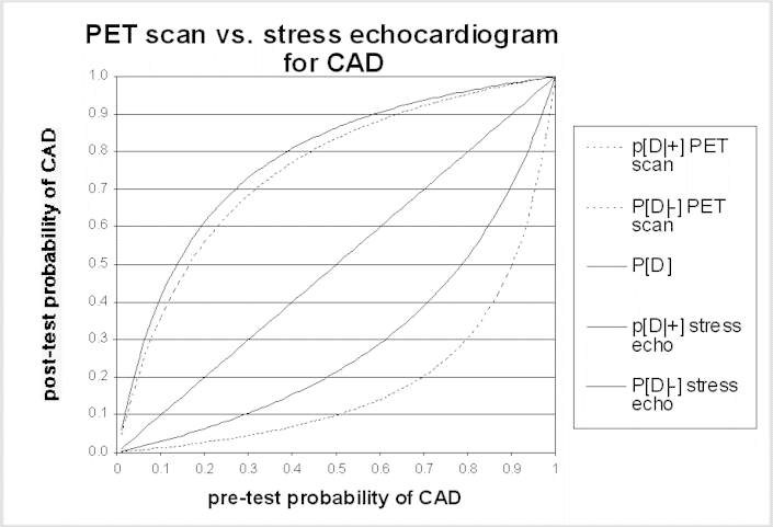
The same principles apply to the use of testing to stage disease or to monitor the effect of treatment. In these situations, the uncertainty is not about the diagnosis, but the test is needed to reduce uncertainty about the current status of the disease. Learning more about stage or response to treatment is important insofar as it will influence management options – for example, disease progression while on one treatment will often lead to a change in therapies, or cessation of a potentially toxic therapy. A false-negative staging test result (i.e., one that implies the disease is more limited than it really is) may lead to treatment that is both ineffective and harmful. In some situations, a false-positive staging test result can have even more harmful consequences; the physician could withhold potentially curative treatment if he or she interprets the staging test as indicating that cure is not possible, dooming a patient to die of a disease that could have been treated effectively.
Step 2: Evaluate the potential impact on management when tests differ in the post-test probability:
In the absence of direct evidence of the effects of a test on health outcomes, it will sometimes be possible to conclude with great confidence that improved accuracy will lead to better outcomes. This is particularly likely to be true when the treatment or management strategy is effective for patients with the disease, but poses risks or discomfort that would not be acceptable when administered to patients who do not have the disease. Then, improved accuracy leads to effective treatment for more people who truly have the disease, and helps avoid unnecessary treatment in people who would not benefit from it. Thus, although the evidence that diagnostic tests for cancer and for heart disease alter health outcomes is largely indirect, it is also compelling. For these categories of disease, there is often strong evidence that treatments with significant adverse consequences are effective when used appropriately. Panels will need to judge whether the test leads to better patient management by increasing the rate at which patients with disease receive appropriate treatment and the rate at which patients who do not have the disease avoid unnecessary treatment.
If management changes, the improvement in health outcomes should be large enough that the panel believes it is clinically significant. A small increase in accuracy can lead to substantial improvements in health outcomes if treatment is highly effective. Improved accuracy is of little consequence, however, if treatment is either ineffective, so there is little benefit to patients with the disease, or very safe, so there is little harm to patients without the disease. Then improved accuracy is unlikely to lead to improved health outcomes or even to influence clinical NCDs.
Under exceptional circumstances, prognostic information, even if it did not affect a treatment decision, could be considered to improve health outcomes. The panel should be alert for circumstances in which patients would be likely to value the prognostic information enough to significantly alter their well-being.
Summary
The recommended approach for evaluating diagnostic tests is as follows:
- Review, when available, high quality studies that provide direct evidence that test results improve health outcomes.
- If there is no high quality direct evidence, evaluate the indirect evidence as follows:
Decide whether studies of test accuracy are sufficiently free of bias to permit conclusions about the accuracy of the test under consideration, in comparison either to another test or another screening, diagnostic, or staging strategy
Evaluate the potential impact of improved accuracy (or complementary information) on health outcomes. Evaluating the effect of test accuracy on post-test probability is one part of this step. The other part is deciding whether the change in patient management that results from the test will improve health outcomes. Improved outcomes are likely to occur when the management strategy is effective in patients with the disease and does not benefit those without the disease. A test can also improve health outcomes when the treatment poses significant risk, so that it is very important to avoid unnecessary treatment.
1The more technical expression of this condition is that a more accurate test is one whose receiver operating characteristic (ROC) curve is above and to the left of the ROC curve for the alternative test.
APPENDIX: THE COMPLEMENTARY VALUE OF COMBINED TESTING
To test the hypothesis that two tests are complementary, several approaches are possible. The best way is a study in which a series of patients receive both tests as well as the reference test. The analysis compares the sensitivity of the second test in two groups of patients: those with a negative result on the first test and those with a positive result, as shown in the table.
| |
Test 1 results positive |
Test 1 results negative |
| Test 2 results |
Reference standard positive |
Reference standard negative |
Reference standard positive |
Reference standard negative |
| Positive |
A |
|
A' |
|
| Negative |
B |
|
B' |
|
| Totals |
A+B |
|
A'+B' |
|
If the sensitivity of Test 2 when test 1 is negative (A'/[A'+B']) is greater than zero, Test 2 is able to detect patients that Test 1 cannot, and the two tests are complementary. If, on the other hand, the sensitivity of Test 2 is zero when Test 1 is negative, Test 2 is unable to detect patients that Test 1 would miss, and it is of minimal additional value.
Many studies of two tests do not provide the information in this table. However, the studies may still provide useful data that reflect what is in the table. The best way to think about using two tests is to consider them as a sequence of tests, in which the post-test probability after the first test becomes the pre-test probability for the second test. Suppose that the test under consideration is the second test in the sequence. It would add information when compared to the established test alone under two circumstances:
- The first test in the sequence is positive, and the post-test probability after a positive result on the second test in the sequence is greater than the post-test probability after the first test.
- The first test in the sequence is negative, and the post-test probability after a negative result on the second test in the sequence is lower than the post-test probability after the first test.
Arguments that consist largely of inductive reasoning (based upon a different physiological basis for Test 2) are much weaker than empirical evidence.


